. 2022 Feb;13(2):2720-2731.
doi: 10.1080/21655979.2021.2001925.

Glycyrrhizic acid exhibits strong anticancer activity in colorectal cancer cells via SIRT3 inhibition

Affiliations

Glycyrrhizic acid exhibits strong anticancer activity in colorectal cancer cells via SIRT3 inhibition

Zhenkui Zuo et al. Bioengineered. 2022 Feb.

Abstract

Sirtuin-3 (SIRT3) has been described as a colorectal cancer oncogene and to be regulated by glycyrrhizic acid (GA). However, few studies have explored the interaction between GA and SIRT3. Therefore, in the present study, we showed that GA could significantly decrease SIRT3 protein levels in SW620 and HT29 cells in a dose-dependent manner. Then, we overexpressed SIRT3 by lentivirus infection on SW620 and HT29 cells. We found that, in vitro, GA treatment significantly decreased cell viability, cell clone number, and invasion and migration number, besides significantly increasing apoptosis. Also, GA treatment significantly decreased the Bax/Bcl2 protein ratio and the expression of Cyclin D1, CDK2, CDK4, MMP-9, N-cadherin, and vimentin in SW620 and HT29 cells. Meanwhile, the SIRT3 overexpression could significantly reverse these changes. Moreover, the GA treatment could significantly decrease the weight of xenograft tumor tissues and its SIRT3 protein levels in vivo, while SIRT3 overexpression reversed these effects. Overall, GA inhibited the proliferation, invasion, and migration of colorectal cancer cells, and induced their apoptosis by SIRT3 inhibition.

Keywords: Glycyrrhizic acid; SIRT3; anticancer; apoptosis; colorectal cancer.


Conflict of interest statement

No potential conflict of interest was reported by the author(s).

Figures

Figure 1.
Figure 1.
Molecular formula of glycyrrhizic acid.
Figure 2.
Figure 2.
Glycyrrhizic acid inhibits SIRT3 protein expression in colorectal cancer cells. (a,b) Western blot analysis of SIRT3 protein expression in SW620 (a) and HT29 (b) cells after stimulating with 0, 5, 10 or 20 μmol/L glycyrrhizic acid for 24 hours. (c,d) Immunofluorescence analysis shows that SIRT3 is mainly expressed in the cytoplasm of SW620 (c) and HT29 (d) cells, and decreased expression of SIRT3 protein after stimulating with 20 μmol/L glycyrrhizic acid for 24 hours. Each test is repeated at least 3 times independently. * P < 0.05, ** P < 0.01 and *** P < 0.001 vs 0 μmol/L glycyrrhizic acid group.
Figure 3.
Figure 3.
Glycyrrhizic acid inhibits the proliferation of colorectal cancer cells by inhibiting SIRT3 in vitro. (a) Lentiviral infection increases the expression of SIRT3 protein in colorectal cancer cells using Western blot analysis. (b) SIRT3 overexpression increases decreased the cell viability of colorectal cancer induced by 20 μmol/L glycyrrhizic acid for 24 hours. (c,d) Wild-type and SIRT3 overexpressing colorectal cancer cells were cultured with 0 or 20 μmol/L glycyrrhizic acid for 2 weeks, representative cell clone images (c), and statistical comparison of the number of cell clones (d). Each test is repeated at least 3 times independently. ns P > 0.05 and *** P < 0.001 vs WT group, and ### P < 0.001 vs 20 μM GA group.
Figure 4.
Figure 4.
Glycyrrhizic acid blocks colorectal cancer cells in G1/G0 stage by inhibiting SIRT3 in vitro. (a,b) Flow cytometry analysis of cell cycle in SW620 (a) and HT29 (b) cells, Representative cell cycle diagrams are shown on the left, and statistical comparison cubes are shown on the right. (c,d) Western blot analysis of Cyclin D1, CDK2 and CDK4 protein expression in SW620 (c) and HT29 (d) cells after stimulating with 0 or 20 μmol/L glycyrrhizic acid for 24 hours. Each test is repeated at least 3 times independently. ** P < 0.01 and *** P < 0.001 vs WT group. # P < 0.05, ## P < 0.01 and ### P < 0.001 vs 20 μM GA group.
Figure 5.
Figure 5.
Glycyrrhizic acid inhibits the metastasis of colorectal cancer cells by inhibiting SIRT3 in vitro. (a,b) Transwell chamber was used to assess the invasion and migration ability of SW620 (a) and HT29 (b) cells. (c,d) Western blot analysis of MMP-9, N-cadherin and Vimentin protein expression in SW620 (c) and HT29 (d) cells after stimulating with 0 or 20 μmol/L glycyrrhizic acid for 24 hours. Each test is repeated at least 3 times independently. *** P < 0.001 vs WT group. ## P < 0.01 and ### P < 0.001 vs 20 μM GA group.
Figure 6.
Figure 6.
Glycyrrhizic acid induces the apoptosis of colorectal cancer cells by inhibiting SIRT3 in vitro. (a,b) 0 or 20 μmol/L glycyrrhizin stimulated colorectal cancer cells for 24 hours, flow cytometry was used to detect the apoptosis of colorectal cancer cells (a) and statistical comparison (b). (c) Western blot analysis of Bax and Bcl2 protein expression in colorectal cancer cells after stimulating with 0 or 20 μmol/L glycyrrhizic acid for 24 hours. (d) Elisa kit was used to determine the activity of caspase 3 in colorectal cancer cells after stimulating with 0 or 20 μmol/L glycyrrhizic acid for 24 hours. Each test is repeated at least 3 times independently. *** P < 0.001 vs WT group, and ### P < 0.001 vs 20 μM GA group.
Figure 7.
Figure 7.
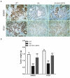
Glycyrrhizic acid inhibits SITR3 expression and the proliferation of colorectal cancer cells in nude mouse. (a-b) Subcutaneous injection of colorectal cancer cells to establish xenograft tumor models in nude mice. 3 weeks later, nude mice were euthanized, xenograft tumors were isolated, SIRT3 protein expression was detected by immunohistochemistry (a), and xenograft tumors were weighed for comparison (b). 8 mice per group. *** P < 0.001 vs WT group, and ### P < 0.001 vs 20 μM GA group.

References

    1. Siegel RL, Miller KD, Fuchs HE, et al. Cancer statistics, 2021. CA Cancer J Clin. 2021;71:7-33. - PubMed
    1. Feng RM, Zong YN, Cao SM, et al. Current cancer situation in China: good or bad news from the 2018 global cancer statistics? Cancer Commun. 2019;39:1–12. - PMC - PubMed
    1. Chen W, Zheng R, Baade PD, et al. Cancer statistics in China, 2015. CA Cancer J Clin. 2016;66:115–132. - PubMed
    1. Zhang Y, Chen Z, Li J.. The current status of treatment for colorectal cancer in China: a systematic review. Medicine. 2017;96:1-6. - PMC - PubMed
    1. Huang HY, Shi JF, Guo LW, et al. Expenditure and financial burden for the diagnosis and treatment of colorectal cancer in China: a hospital‐based, multicenter, cross‐sectional survey. Cancer Commun. 2017;36:1–15. - PMC - PubMed

Publication types

MeSH terms