Review
. 2021 Jan-Dec;13(1):1990827.
doi: 10.1080/19490976.2021.1990827.

Gut microbiota: sculptors of the intestinal stem cell niche in health and inflammatory bowel disease

Affiliations
Review

Gut microbiota: sculptors of the intestinal stem cell niche in health and inflammatory bowel disease

Manasvini Markandey et al. Gut Microbes. 2021 Jan-Dec.

Abstract

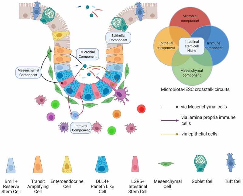
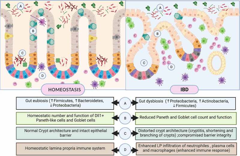
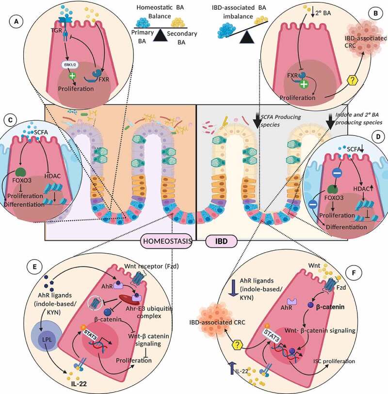
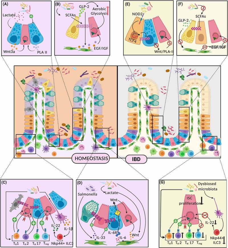
Intestinal epithelium represents a dynamic and diverse cellular system that continuously interacts with gut commensals and external cues. Intestinal stem cells, which lie at the heart of epithelial renewal and turnover, proliferate to maintain a steady stem cell population and differentiate to form functional epithelial cell types. This rather sophisticated assembly-line is maintained by an elaborate micro-environment, sculpted by a myriad of host and gut microbiota-derived signals, forming an intestinal stem cell niche. This complex, yet crucial signaling niche undergoes dynamic changes during homeostasis and chronic intestinal inflammation. Inflammatory bowel disease refers to a chronic inflammatory response toward pathogenic or commensal microbiota, in a genetically susceptible host. Compositional and functional alterations in gut microbiota are pathognomonic of IBD.The present review highlights the modulatory role of gut microbiota on the intestinal stem cell niche during homeostasis and inflammatory bowel disease. We discuss the mechanisms of direct action of gut commensals (through microbiota-derived or microbiota-influenced metabolites) on ISCs, followed by their effects via other epithelial and immune cell types.

Keywords: Intestinal epithelial stem cells (ISCs); crypt specific core microbiota CSCM); enteroendocrine cells; gut metabolites; gut microbiota; inflammatory bowel disease (IBD); mesenchyme; paneth cells.


Conflict of interest statement

No potential conflict of interest was reported by the author(s).

Figures

Figure 1.
Figure 1.
The components of the intestinal stem cell niche. The intestinal stem cell niche, which lies at the heart of gut epithelial renewal and regeneration, constitutes the factors contributed by four intestinal cellular compartments – the gut microbiota, the epithelium, immune cells of the underlying lamina propria and the mesenchymal cells. The gut microbiota regulates ISC physiology either directly or through other niche components, shaping the epithelial renewal during homeostasis and post-injury regeneration
Figure 2.
Figure 2.
The intestinal stem cell niche along colon in homeostasis (left panel) and Inflammatory Bowel Disease (right panel). Intestinal stem cell niche comprises of intestinal stem cells and Paneth-like population of Dll4+/Reg4+ Deep Crypt Cells (DCS) located along the crypt base. ISCs undergoes continual renewal and differentiation into distinct secretory and absorptive epithelial cell types. Other sub-niches such as the gut microbiota and underlying layer of immune cells, contribute to the myriad of signals which drive this complex and dynamic process of epithelial renewal and differentiation. IBD-associated alterations in ISC niche and the associated factors have been highlighted along the lower panel
Figure 3.
Figure 3.
Interaction of Gut Microbiota-derived or influenced metabolites on ISC niche during homeostasis (left panel) and IBD (right panel); Homeostatic levels of primary and secondary bile acid pools, through membrane-bound TGR and cytosolic FXR, is critical for maintaining ISC proliferation (a). Deterioration of microbiota producing the secondary BAs during IBD, promotes uncontrolled ISC proliferation (b). SCFAs modulate ISC physiology through transcriptional and epigenetic regulation (c). IBD- associated dwindling levels of SCFAs, disturbs these regulatory pathways (d). Microbiota-derived AhR ligands checks the ISC proliferation through degradation of β-catenin, by ubiquitination and preventing the cell surface expression of Wnt ligands on ISCs (e) The loss of these regulatory pathways due to IBD-associated degradation of gut indole pool, may drive uncontrolled ISC proliferation (f). TRG (G-protein coupled bile acid receptor), BA (Bile acid), FXR (Farnesoid X Receptor), SCFA (Short-chain fatty acids), HDACs (Histone deacetylases), KYN (Kynurenine), AhR (Aryl-hydrocarbon receptor), Fzd (Frizzled)
Figure 4.
Figure 4.
Indirect mechanisms of interaction between Gut Microbiota and LGR5+ Intestinal Stem Cells. (Figure 4 follows the key attached with Figure 3 for cell type reference) Significant interactions along the gut microbiota – intestinal stem cell axis, and their effects on proliferation and differentiation of ISCs have been depicted in homeostasis (left panel) and IBD (right panel). Paneth cells mediate these interactions through release of ISC proliferative signals like Wnt3A and phospholipase A2 (PLA II) (a). During IBD, these PC-mediated ISC-microbiota interactions are dysregulated (e). Enteroendocrine cells (EECs) forge microbiota-ISC interactions through the release of GLP-2 in response to microbial metabolites like SCFAs, which not only regulates their own differentiation from progenitors, but also drives underlying mesenchyme to release growth factors supporting ISC renewal (b). IBD – characterized dysbiosed microbiota leads to alterations in these interaction (f). Microbial stimulation can similarly lead to different helper T lymphocyte populations (Th1, Th2, Th17 and Treg) and ILC3, mediating either ISC self-renewal or differentiation (c). These mechanisms go haywire during IBD (g). Mesenchymal component of the ISC niche produces factors like IL-33, IL-6 and Wnt, in response to microbial signals, which regulate ISC proliferation and differentiation (d). PLA II (Phospholipase A2), SCFA (Short-chain fatty acids), GLP-2 (Glucagon-like peptide 2), EGF (Epidermal growth factor)/IGF (Insulin-like growth factor)

References

    1. Barker N, van Es JH, Kuipers J, Kujala P, van den Born M, Cozijnsen M, Haegebarth A, Korving J, Begthel H, Peters PJ, et al. Identification of stem cells in small intestine and colon by marker gene Lgr5. Nature. 2007; 449:1003–21. doi:10.1038/nature06196 - DOI - PubMed
    1. Sato T, Vries RG, Snippert HJ, van de Wetering M, Barker N, Stange DE, van Es JH, Abo A, Kujala P, Peters PJ, et al. Single Lgr5 stem cells build crypt-villus structures in vitro without a mesenchymal niche. Nature. 2009; 459:262–5. doi:10.1038/nature07935 - DOI - PubMed
    1. Saffarian A, Mulet C, Regnault B, Amiot A, Tran-Van-Nhieu J, Ravel J, Sobhani I, Sansonetti PJ, Pédron T. Crypt- and Mucosa-Associated Core Microbiotas in Humans and Their Alteration in Colon Cancer Patients. MBio. 2019; 10. doi:10.1128/mBio.01315-19 - DOI - PMC - PubMed
    1. Pédron T, Mulet C, Dauga C, Frangeul L, Chervaux C, Grompone G, Sansonetti PJ. A crypt-specific core microbiota resides in the mouse colon. MBio. 2012; 3. doi:10.1128/mBio.00116-12 - DOI - PMC - PubMed
    1. Guan Q. A Comprehensive Review and Update on the Pathogenesis of Inflammatory Bowel Disease. J Immunol Res. 2019. doi:10.1155/2019/7247238. - DOI - PMC - PubMed

Publication types

MeSH terms

LinkOut - more resources