. 2021 Nov 16;118(46):e2111451118.
doi: 10.1073/pnas.2111451118.

Mutation in Abl kinase with altered drug-binding kinetics indicates a novel mechanism of imatinib resistance

Affiliations

Mutation in Abl kinase with altered drug-binding kinetics indicates a novel mechanism of imatinib resistance

Agatha Lyczek et al. Proc Natl Acad Sci U S A. .

Abstract

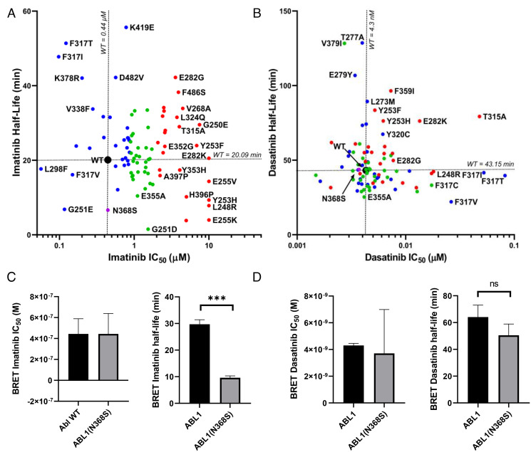
Protein kinase inhibitors are potent anticancer therapeutics. For example, the Bcr-Abl kinase inhibitor imatinib decreases mortality for chronic myeloid leukemia by 80%, but 22 to 41% of patients acquire resistance to imatinib. About 70% of relapsed patients harbor mutations in the Bcr-Abl kinase domain, where more than a hundred different mutations have been identified. Some mutations are located near the imatinib-binding site and cause resistance through altered interactions with the drug. However, many resistance mutations are located far from the drug-binding site, and it remains unclear how these mutations confer resistance. Additionally, earlier studies on small sets of patient-derived imatinib resistance mutations indicated that some of these mutant proteins were in fact sensitive to imatinib in cellular and biochemical studies. Here, we surveyed the resistance of 94 patient-derived Abl kinase domain mutations annotated as disease relevant or resistance causing using an engagement assay in live cells. We found that only two-thirds of mutations weaken imatinib affinity by more than twofold compared to Abl wild type. Surprisingly, one-third of mutations in the Abl kinase domain still remain sensitive to imatinib and bind with similar or higher affinity than wild type. Intriguingly, we identified three clinical Abl mutations that bind imatinib with wild type-like affinity but dissociate from imatinib considerably faster. Given the relevance of residence time for drug efficacy, mutations that alter binding kinetics could cause resistance in the nonequilibrium environment of the body where drug export and clearance play critical roles.

Keywords: binding kinetics; drug binding; drug resistance; imatinib; protein kinase.


Figures

Fig. 1.
Fig. 1.
In-cell inhibitor–binding affinities of mutant panel. (A) Structure of the Abl KD (PDB entry 2HYY). The locations of 94 patient-derived Abl mutants associated with resistance are indicated as spheres. The color of the spheres indicates the level of imatinib resistance conferred by the mutation: blue, affinity of mutant is less than twofold weaker than wt; green, affinity is between two- and fourfold weaker than wt; red, affinity is more than eightfold weaker than wt. (B) Fraction of mutations in different resistance classes. The colors correspond to A. (C) Imatinib and dasatinib resistance distribution of Abl mutants determined by in-cell NanoBRET target engagement assay. Colors correspond to A. Confidence of values is ± 23% based off of wt SD, n = 2.
Fig. 2.
Fig. 2.
In-cell dissociation kinetics. (A and B) Abl mutants were characterized for imatinib (A) and dasatinib (B) affinity and dissociation kinetics using NanoBRET assay in live cells. Mutants were colored according to imatinib affinity as in Fig. 1. The novel Abl N368S mutant was characterized for (C) imatinib- and (D) dasatinib-binding affinities and dissociation kinetics by NanoBRET in live cells. ns: P > 0.05, ***P < 0.001.
Fig. 3.
Fig. 3.
Characterization of the Abl N368S mutation. (A) Sequence alignment of human protein kinases represented as weblogo. The size of the letters reflects the degree of conservation, and numbering is according to Abl1A numbering. Asparagine 368 is as highly conserved as the catalytic His-Arg-Asp (HRD) and the DFG motif. (B) Structural overview of Abl N368 in the active conformation (PDB entry 2G2I). The sidechain of N368 is near the nucleotide and is positioned to form hydrogen bonds with the catalytic aspartate D363 and D381 in the DFG motif. (C) Kinase activity of purified Abl N368S is undetectable in the absence of activation loop phosphorylation (designated as pAbl). (D) The Michaelis–Menten constant for ATP of purified pAbl N368S is twofold higher than that of pAbl wt. (E) The affinity of purified Abl N368S for imatinib determined by a spectroscopic-binding assay is slightly weaker than for Abl wt. (F) BaF3 cells expressing Bcr-Abl wt, T315I, or N368S. BaF3 cells expressing N368S are equally sensitive to imatinib as cells expressing Bcr-Abl wt. (G) BaF3 cells expressing imatinib resistance mutations T315I or N368S exhibit a faster doubling time in the absence of imatinib. ns: P > 0.05, **P < 0.01, ***P < 0.001, ****P < 0.0001.
Fig. 4.
Fig. 4.
In vitro kinetics of imatinib binding and dissociation from purified Abl KD. (A and B) Dissociation kinetics of imatinib from unphosphorylated (A) and activation loop–phosphorylated (B) Abl wt and N368S at various temperatures. (C and D) Dissociation of imatinib from unphosphorylated (C) and activation loop–phosphorylated (D) Abl wt and N368S at various pH values. (E and F) Binding kinetics of imatinib to Abl wt and N368S at various pH values yielding the rate constant of imatinib binding (E) and the rate constant of the DFG-flip (F).
Fig. 5.
Fig. 5.
MD and NMR analyses of the N368S mutant protein. (A) Fraction of snapshots from MD simulation in which the sidechain of N368 engages in a hydrogen bond with neighboring atoms (Left) illustrated in the structure of Abl kinase in the active conformation (PDB entry 2GQG) (Right). (B) Fraction of snapshots from MD simulations in which the sidechain of S368 engages with neighboring atoms (Left) illustrated on the model of S368 in the structure of active Abl wt (PDB entry 2GQG). (C) NMR 1H-15N peak intensity ratios (IN368S/IWT) for residues in helix αC and the activation loop. Black line: ratio of 1, yellow line: mean of all values larger than 1, red line: 1 SD from mean of values larger than 1, cyan line: mean of all values smaller than 1, purple line: 1 SD from mean of values smaller than 1.

Similar articles

Cited by

References

    1. Jemal A., et al. ., Cancer statistics, 2007. CA Cancer J. Clin. 57, 43–66 (2007). - PubMed
    1. Parker S. L., Tong T., Bolden S., Wingo P. A., Cancer statistics, 1997. CA Cancer J. Clin. 47, 5–27 (1997). - PubMed
    1. Gorre M. E., et al. ., Clinical resistance to STI-571 cancer therapy caused by BCR-ABL gene mutation or amplification. Science 293, 876–880 (2001). - PubMed
    1. Azam M., Latek R. R., Daley G. Q., Mechanisms of autoinhibition and STI-571/imatinib resistance revealed by mutagenesis of BCR-ABL. Cell 112, 831–843 (2003). - PubMed
    1. Shah N. P., et al. ., Multiple BCR-ABL kinase domain mutations confer polyclonal resistance to the tyrosine kinase inhibitor imatinib (STI571) in chronic phase and blast crisis chronic myeloid leukemia. Cancer Cell 2, 117–125 (2002). - PubMed

Publication types

MeSH terms