. 2021 Nov 8;12(1):6447.
doi: 10.1038/s41467-021-26531-1.

Nascent chains can form co-translational folding intermediates that promote post-translational folding outcomes in a disease-causing protein

Affiliations

Nascent chains can form co-translational folding intermediates that promote post-translational folding outcomes in a disease-causing protein

Elena Plessa et al. Nat Commun. .

Abstract

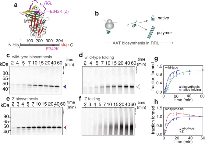
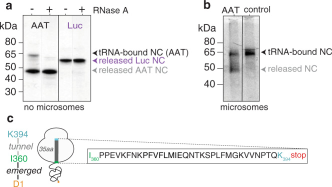
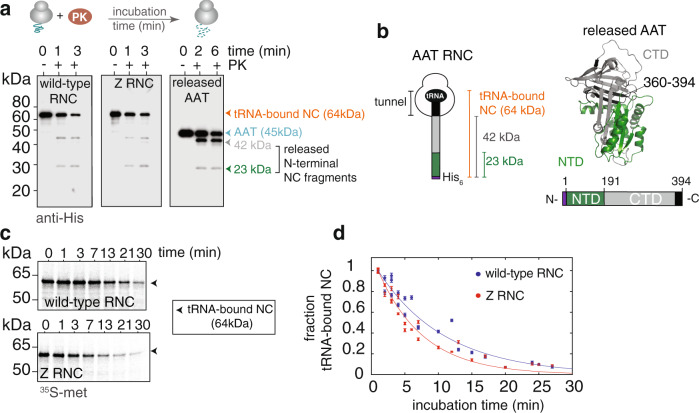
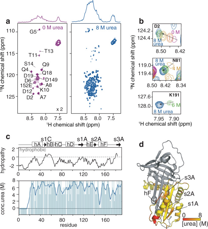
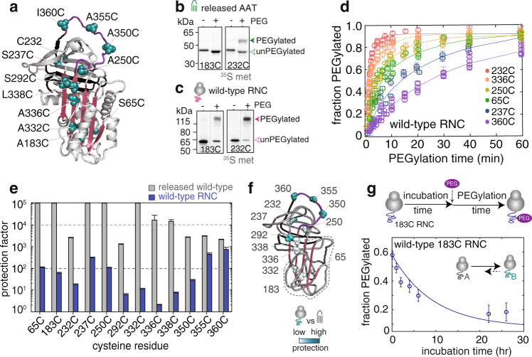
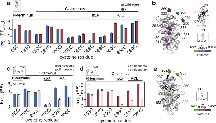
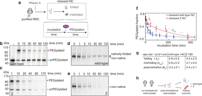
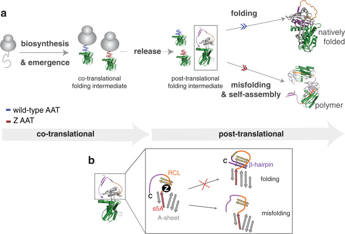
During biosynthesis, proteins can begin folding co-translationally to acquire their biologically-active structures. Folding, however, is an imperfect process and in many cases misfolding results in disease. Less is understood of how misfolding begins during biosynthesis. The human protein, alpha-1-antitrypsin (AAT) folds under kinetic control via a folding intermediate; its pathological variants readily form self-associated polymers at the site of synthesis, leading to alpha-1-antitrypsin deficiency. We observe that AAT nascent polypeptides stall during their biosynthesis, resulting in full-length nascent chains that remain bound to ribosome, forming a persistent ribosome-nascent chain complex (RNC) prior to release. We analyse the structure of these RNCs, which reveals compacted, partially-folded co-translational folding intermediates possessing molten-globule characteristics. We find that the highly-polymerogenic mutant, Z AAT, forms a distinct co-translational folding intermediate relative to wild-type. Its very modest structural differences suggests that the ribosome uniquely tempers the impact of deleterious mutations during nascent chain emergence. Following nascent chain release however, these co-translational folding intermediates guide post-translational folding outcomes thus suggesting that Z's misfolding is initiated from co-translational structure. Our findings demonstrate that co-translational folding intermediates drive how some proteins fold under kinetic control, and may thus also serve as tractable therapeutic targets for human disease.


Conflict of interest statement

The authors declare no competing interests.

Figures

Fig. 1
Fig. 1. Monitoring the folding and misfolding of alpha-1-antitrypsin (AAT) during biosynthesis in rabbit reticulocyte lysate (RRL) using 35S methionine radiolabelling.
a (upper) AAT structure (PDB ID: 1QLP) with landmarks highlighted: A-sheet (red), B-sheet (green), C-sheet (yellow), reactive centre loop (RCL, purple), and site of the Z mutation (E342K). (lower) Schematic of the full-length AAT DNA construct and highlighting the relative position of the Z (E342K) mutation. b Scheme of released AAT biosynthesis and folding in RRL. c Production of wild-type AAT nascent chains as followed by denaturing PAGE. Highlighted are the full-length species (arrow), and high molecular weight species / polymerisation (i.e., a smear) as a dashed line. d Folding of wild-type AAT nascent chains as monitored by native PAGE (using the same samples as in c). Highlighted is the biologically-active, monomeric species (arrow) and polymer (dashed line). e Production of Z nascent chains as followed by denaturing PAGE. Highlighted are the full-length species (arrow), and high molecular weight species / polymerisation (i.e., a smear) as a dashed line. f Folding of Z AAT nascent chains as monitored by native PAGE (using the same samples as in e). Unlike wild-type in c, Z nascent chains cannot fold efficiently (solid line) and form polymers (dashed line). g Biosynthesis and native folding of wild-type AAT nascent chains (analysis from c, d; n = 4). h Biosynthesis of wild-type and Z AAT nascent chains over 60 min. Data have been normalised to the reaction end-point to enable a comparative analysis. All samples were flash-frozen and treated with RNase A prior to analysis. (n = 5 biological repeats). Data are presented as mean values +/− SEM.
Fig. 2
Fig. 2. AAT forms persistent full-length ribosome-nascent chain complexes.
a Biosynthesis of AAT and luciferase (Luc) after a 60 min biosynthesis reaction quenched with cycloheximide in the absence of microsomes (both DNA constructs contain stop codons). Highlighted are the full-length, tRNA-bound nascent chains (NC) and full-length, released nascent chains. b Biosynthesis of AAT in the presence of microsomes, alongside a control (AAT DNA construct with no stop codon to force production of RNCs), highlighting that both full-length tRNA-bound nascent chains (NC) and released nascent chains can be formed in the endoplasmic reticulum. Source data are provided as a Source Data file. c Schematic of a full-length AAT RNC showing the sequence expected within the tunnel and beyond the ribosome. Note that a and b show 35S methionine detection via partially-denaturing PAGE (see “Methods” section).
Fig. 3
Fig. 3. Wild-type and Z AAT ribosome-nascent chain complexes (RNC) can adopt co-translational structure.
a A schematic of the proteinase K limited proteolysis experiment (upper). Limited proteolysis of wild-type and Z purified RNCs (orange arrowhead), and purified, released wild-type AAT (blue arrowhead) monitored by an anti-His western blot (lower). Highlighted are two N-terminal proteolytic products released from the nascent chain segment emerged from the ribosome: 42 kDa (grey arrowhead) and 23 kDa (green arrowhead). b Schematic of an AAT RNC highlighting the N-terminal fragments observed in a (left). (right) Structure of AAT and schematic representation of (released) AAT showing the boundaries of the N-terminal (NTD, green) and C-terminal (CTD, grey) sub-domains, and C-terminal residues 360–394 which are occluded in the ribosomal tunnel within the RNC (see left). The segment of structure shown in black is the 35 amino acid stretch that is occluded in the tunnel when full-length AAT is bound to the ribosome. c Proteinase K limited proteolysis measured over time for wild-type and Z RNCs, highlighting the measurement of the intact tRNA-bound nascent chain (NC) over time using 35S-methionine detection and partially-denaturing PAGE (n = 3 biological repeats). See also Supplementary Fig. 2. d Densitometric analysis of the proteinase K time-course from c with exponential fits. Data are presented as mean values +/− SEM.
Fig. 4
Fig. 4. NMR characterisation of the N-terminal fragment AAT-191.
a (upper) 1D and (lower) 2D 1H,15N-SOFAST HMQC spectra of the AAT-191 fragment in (left) 0 M and (right) 8 M urea, recorded at 25 °C and at a 1H frequency of 950 MHz. b Selected AAT-191 resonances observed in increasing concentrations of urea. c (upper) A Kyte and Doolittle hydropathy plot for AAT-191 (lower) A plot depicting the urea concentrations at which non-proline residues are first observable, and includes a 3-point moving average to highlight the extent of compaction observed across the sequence. d Mapped onto the AAT structure is a representation of AAT-191 depicting the regions that progressively unfold in 8 M urea and at which stage NMR resonances first become observable (c).
Fig. 5
Fig. 5. Full-length, ribosome-bound AAT nascent chains form a co-translational folding intermediate.
a Structure of AAT (1QLP) with all single cysteine probe positions highlighted. b Extent of PEGylation in cysteine variants 183C and 232C in released, natively-folded wild-type AAT measured after a 60 min PEGylation reaction at 25 °C. See also Supplementary Fig. 4). c As described for b, but shows wild-type ribosome-nascent chain complexes (RNC). Source data are provided as a Source Data file. d Fitted PEGylation kinetics of selected wild-type RNCs. For clarity, only the first 60 min (of 180 min) are shown (n = 4). See Supplementary Fig. 4. e Comparison of protection factors of released wild-type AAT and wild-type RNCs at 25 °C. Shown in the dashed lines are the average protection factor values for released wild-type and wild-type RNCs (n = 5 biological repeats). f Structural depiction of the co-translational folding intermediate of wild-type, derived from a comparison of protections factors against released, natively-folded wild-type AAT. Highlighted in dashed lines is the N-terminal fragment, 1–191 (analysis taken from e). g (upper) Schematic of a PEGylation reaction for wild-type AAT RNCs highlighting an (RNC) incubation time prior to PEGylation. (lower) Extent of PEGylation for wild-type 183C RNCs as measured across different RNC incubation times. Each data point is a different RNC incubation time-point. (inset) a kinetic model for RNC behaviour as observed over time. (n = 5 biological repeats). Data are presented as mean values +/− SEM.
Fig. 6
Fig. 6. Co-translational folding intermediates persist post-translationally.
a Protection factors (PF) calculated for the co-translational folding intermediates formed by wild-type and Z RNCs as derived from PEGylation kinetics (n = 5 biological repeats). b Difference in protection in the Z’s co-translational folding intermediate relative to that of wild-type, as mapped onto AAT’s native structure. (inset) A magnified view of AAT’s central A-sheet region, highlighting the location of the s5A/RCL region and the Z mutation. This region is implicated in AAT folding (see text). c A comparison of the protection factors for wild-type’s co-translational folding intermediate (on ribosome) and post-translational folding intermediate (off ribosome) for a series of cysteine positions. d As described for c but shows Z. e Difference in protection in Z’s post-translational intermediate relative to that of wild-type, as mapped onto AAT’s native structure. Data are presented as mean values +/− SEM.
Fig. 7
Fig. 7. Monitoring post-translational folding outcomes of released AAT nascent chains (NC).
a A reaction scheme for monitoring post-translational folding by PEGylation, in which nascent chains are synchronously-released from 183C RNCs using RNase A to initiate their (off the ribosome) folding. PEG is added at intervals to monitor the folding process, and the PEGylation reaction time is 60 min at 25 °C. (n = 5 biological repeats). b PEGylation of RNase A-released nascent chains of wild-type 183C monitored via partially-denaturing PAGE. c As described for b, but shows Z 183C. d Folding of RNase A-released, wild-type nascent chains as monitored by native PAGE (equivalent samples as shown in (b), but without PEGylation). e Folding of RNaseA-released, Z nascent chains as monitored by native PAGE (equivalent samples as shown in c, but without PEGylation). f A PEGylation profile for RNAse A-released wild-type and Z 183C nascent chains from their respective RNCs (analysis of b, c, respectively). Data are presented as mean values +/− SEM. g Rates of post-translational folding, misfolding and polymerisation of RNase A-released 183C nascent chains (n = 4 biological repeats). h A schematic illustrating the tempering effect imposed by ribosome on emerging nascent chains which is removed after the nascent chains are released. All PAGE used 35S methionine detection.
Fig. 8
Fig. 8. Co-translational and post-translational folding and misfolding AAT.
a A folding and misfolding model for AAT during biosynthesis. Highlighted in green is the 1–191 fragment that forms co-translational structure on the ribosome. b A schematic showing how Z’s kinetic folding defect, which begins developing co-translationally, guides post-translational folding outcomes. Shown is a magnified view of the A-sheet/RCL region in the post-translational intermediate: A-sheet (grey, and strand 5A (s5A) in red), B-sheet (gold), reactive centre loop (RCL), orange, β-hairpin (purple). The insertion of the final 35 C-terminal residues to form a β-hairpin in AAT’s core and complete the native fold, is a key component of the folding/misfolding partition.

References

    1. Balchin D, Hayer-Hartl M, Hartl FU. In vivo aspects of protein folding and quality control. Science. 2016;353:aac4354. doi: 10.1126/science.aac4354. - DOI - PubMed
    1. Chiti F, Dobson CM. Protein misfolding, amyloid formation, and human disease: a summary of progress over the last decade. Annu. Rev. Biochem. 2017;86:27–68. doi: 10.1146/annurev-biochem-061516-045115. - DOI - PubMed
    1. Cassaignau AME, Cabrita LD, Christodoulou J. How does the ribosome fold the proteome? Annu. Rev. Biochem. 2020;89:389–415. doi: 10.1146/annurev-biochem-062917-012226. - DOI - PubMed
    1. Waudby, C. A., Dobson, C. M. & Christodoulou, J. Nature and regulation of protein folding on the ribosome. Trends Biochem. Sci. 10.1016/j.tibs.2019.06.008 (2019). - PMC - PubMed
    1. Sato S, Ward CL, Kopito RR. Cotranslational ubiquitination of cystic fibrosis transmembrane conductance regulator in vitro. J. Biol. Chem. 1998;273:7189–7192. doi: 10.1074/jbc.273.13.7189. - DOI - PubMed

Publication types

MeSH terms

Supplementary concepts