. 2022 Mar;36(3):701-711.
doi: 10.1038/s41375-021-01462-4. Epub 2021 Nov 8.

The IL-3, IL-5, and GM-CSF common receptor beta chain mediates oncogenic activity of FLT3-ITD-positive AML

Affiliations

The IL-3, IL-5, and GM-CSF common receptor beta chain mediates oncogenic activity of FLT3-ITD-positive AML

Anne Charlet et al. Leukemia. 2022 Mar.

Abstract

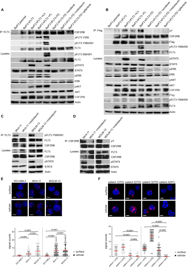
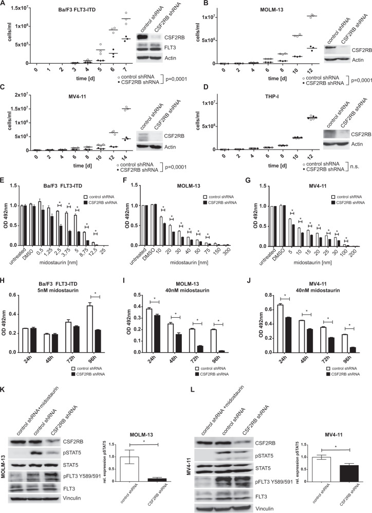
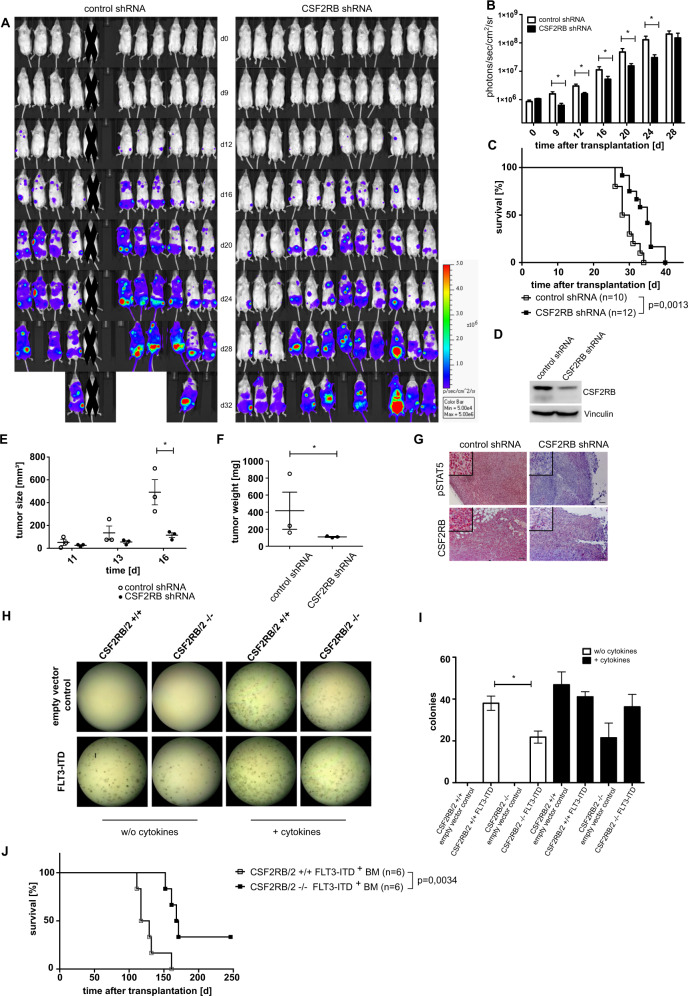
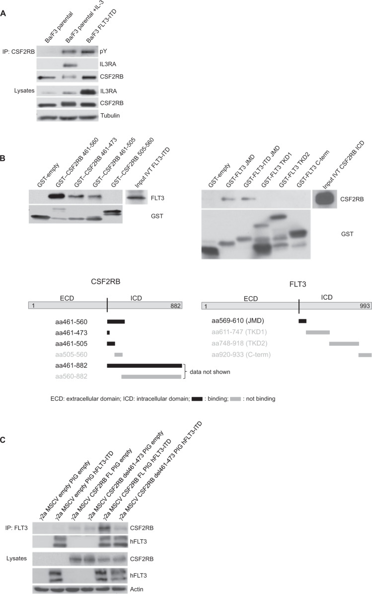
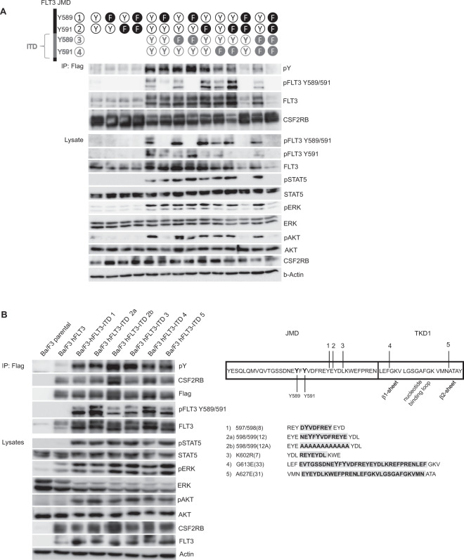
FLT3-ITD is the most predominant mutation in AML being expressed in about one-third of AML patients and is associated with a poor prognosis. Efforts to better understand FLT3-ITD downstream signaling to possibly improve therapy response are needed. We have previously described FLT3-ITD-dependent phosphorylation of CSF2RB, the common receptor beta chain of IL-3, IL-5, and GM-CSF, and therefore examined its significance for FLT3-ITD-dependent oncogenic signaling and transformation. We discovered that FLT3-ITD directly binds to CSF2RB in AML cell lines and blasts isolated from AML patients. A knockdown of CSF2RB in FLT3-ITD positive AML cell lines as well as in a xenograft model decreased STAT5 phosphorylation, attenuated cell proliferation, and sensitized to FLT3 inhibition. Bone marrow from CSF2RB-deficient mice transfected with FLT3-ITD displayed decreased colony formation capacity and delayed disease onset together with increased survival upon transplantation into lethally irradiated mice. FLT3-ITD-dependent CSF2RB phosphorylation required phosphorylation of the FLT3 juxtamembrane domain at tyrosines 589 or 591, whereas the ITD insertion site and sequence were of no relevance. Our results demonstrate that CSF2RB participates in FLT3-ITD-dependent oncogenic signaling and transformation in vitro and in vivo. Thus, CSF2RB constitutes a rational treatment target in FLT3-ITD-positive AML.


Conflict of interest statement

N.v.B. received honoraria from Amgen, Astra Zeneca, BMS, and Novartis, and research funding from Novartis. J.D. received honoraria from Novartis. The authors declare no additional financial interests.

Figures

Fig. 1
Fig. 1. CSF2RB interacts with FLT3.
A, B Ba/F3 cells expressing CSF2RB-Flag and either FLT3 or FLT3-ITD were serum-deprived for 5 h with addition of 2 ng/ml IL-3 (5 min), 50 ng/ml FLT-ligand (5 min), 100 nM midostaurin (1.5 h), 300 nM sorafenib (1,5 h) or 100 nM gilteritinib (1,5 h) as indicated. Co-immunoprecipitations were performed using FLT3-antibody (A) or anti-flag beads capturing CSF2RB (B). Immunoprecipitates and whole-cell lysates were subjected to SDS–PAGE and western blot analysis using indicated antibodies. C, D FLT3-ITD-positive AML cell lines MV4–11 and MOLM-13 were serum-deprived for 4 h and additionally treated with 100 nM midostaurin for 1.5 h as indicated. Co-immunoprecipitations were performed using FLT3-antibody (C) or CSF2RB-antibody (D). Immunoprecipitates and whole-cell lysates were subjected to SDS–PAGE and western blot analysis using indicated antibodies. E Proximity ligation assay (1-PLA) was performed using oligo-coupled primary antibodies against FLT3 and CSF2RB. FLT3 expressing OCI-AML3 cells and FLT3-ITD expressing MV4–11 and MOLM-13 cells were fixed (surface) or fixed and permeabilized with 0.5% saponine (cellular) prior to PLA reaction. Red dots indicate the occurrence of a close FLT3: CSF2RB proximity. Nuclei were counterstained with DAPI. Representative images are shown (Zeiss 780 Meta confocal microscope; Objective NA 1.4), scale bar = 10 µm. Quantification of the PLA signals is shown as signals per cell. F Blast cells from three FLT3-ITD positive AML patients and one FLT3-ITD negative AML patient (patient 4) were isolated from peripheral blood using Ficoll density gradient. Cells were fixed (surface) or fixed and permeabilized (cellular) and PLA was performed (as described in E), p ≤ 0.0002.
Fig. 2
Fig. 2. CSF2RB knockdown impairs FLT3-ITD-induced cell growth, sensitizes cells to FLT3 inhibition, and decreases phosphorylation of STAT5.
AJ Ba/F3 cells transduced with FLT3-ITD, FLT3-ITD-positive AML cell lines MOLM-13 and MV4–11, as well as FLT3 expressing cell line THP-I, were transduced with constructs bearing inducible CSF2RB shRNA or control shRNA, respectively, and cultured in the presence of doxycycline for at least 48 h prior to and during experiments. AD Proliferation of control shRNA vs. CSF2RB shRNA-expressing cells was determined by counting viable cells after trypan blue staining in Neubauer counting chamber at indicated time points. Cells were seeded in triplicates in a density of 1 × 104cells/ml at day 0. Fresh medium was added regularly to maintain optimal cell densities over time. Cell lysates were subjected to SDS–PAGE and western blot analysis using indicated antibodies to confirm knockdown. EG Cell viability was determined using MTS assay. Cells were seeded in a density of 6000 cells per well in 96-well plates and cultured in the presence of midostaurin at the indicated concentrations. Proliferation was measured in triplicates as formazan absorption after 72 h at 490 nM. HJ Cells were seeded in a density of 6000 cells per well in 96-well plates and cultured in the presence of midostaurin at the indicated concentrations. Cell viability was measured daily by formazan absorption at 490 nm. K, L Cells were serum-deprived for 8 h, with or without the addition of 25 nM midostaurin. Whole-cell lysates were subjected to SDS–PAGE and western blot analysis using the indicated antibodies. Quantification of pSTAT5 relative to total STAT5 was calculated from three biologically independent replicates.
Fig. 3
Fig. 3. Loss of CSF2RB attenuates FLT3-ITD-dependent transformation and proliferation and prolongs survival in xenograft and bone marrow transplantation models.
AD Immunodeficient Rag2/Il2rg-mutant mice were sublethally irradiated and injected intravenously with FLT3-ITD-positive MOLM-13 cells stably expressing CSF2RB shRNA (n = 12) or control shRNA (n = 10) together with luciferase. One animal was excluded as no signs of engraftment could be detected at any time point. A Mice were injected intraperitoneally with luciferin and subjected to bioluminescence imaging, as indicated. B Quantification of bioluminescence is shown. P value was calculated using a two-sided Mann–Whitney test. C Survival curve of mice transplanted with MOLM-13 cells expressing CSF2RB shRNA or control shRNA. P values were calculated by Mantel–Cox test, n represents biologically independent mice. D The same MOLM-13 cells were subjected to lysis, SDS–PAGE, and western blot using the indicated antibodies. EG FLT3-ITD positive MOLM-13 cells expressing CSF2RB shRNA or control shRNA were injected subcutaneously into the flank (n = 3 for both groups) of immunodeficient Rag2/Il2rg-mutant mice. E Tumor size was measured at given days by caliper. F Tumor weight was measured after sacrificing the animals on day 17 (p value of 0.0014 calculated by F test). G Sectioned tumors were fixed in 4% paraformaldehyde and then subject to immunohistochemical staining (by labeled Streptavidin–Biotin method) with the indicated antibodies, using hematoxylin for counterstaining. Representative images are shown (Axio Imager.M2; EC Plan-Neofluar ×10/0.3), scale bar = 100 µm. H, I Bone marrow of Csf2rb/Csf2rb2 double-knockout mice and wildtype littermates was transduced with FLT3-ITD or empty vector with GFP as transduction control. In all, 3000 GFP-positive sorted cells were cultured in methylcellulose ± cytokines (SCF, IL-3, IL-6, EPO, insulin, transferrin, iron-saturated). Representative images of one experiment (H) and analysis of three independent experiments (I) are shown. J Lethally radiated C57/BL6 littermates were transplanted with bone marrow from Csf2rb/Csf2rb2 double knockout or wildtype littermates transduced with FLT3-ITD 598/599[12] construct with GFP as transduction control; non-transduced bone marrow was supplemented to equalize the number of GFP-positive cells (n = 6 for both groups). The recipients’ survival is shown, with day 246 after transplantation as an endpoint. P value was calculated by Mantel–Cox test; n represents biologically independent mice.
Fig. 4
Fig. 4. Interaction of CSF2RB and FLT3 is independent of physiological receptor complex formation and takes place at the cytosolic site close to the membrane.
A Parental Ba/F3 cells and FLT3-ITD-expressing Ba/F3 cells were serum-deprived for 4 h and left untreated or incubated with IL-3 at 2 ng/ml. Whole-cell lysates were subjected to immunoprecipitation using antibodies against CSF2RB. Immunoprecipitates and whole-cell lysates were subjected to SDS–PAGE and western blot analysis using indicated antibodies. B Mapping of the CSF2RB and FLT3 binding sites by GST-pulldown assay. Schematic diagrams show the regions used for glutathione S-transferase tagged peptides captured by glutathione-coated agarose beads. In vitro translated intracellular domains of FLT3-ITD and CSF2RB served as binding partners (input). Incubation was performed overnight; interaction complexes were separated by SDS–PAGE and subjected to western blot analysis using the indicated antibodies. JMD juxtamembrane domain, TKD1 tyrosine kinase domain 1, TKD2 tyrosine kinase domain 2. C CSF2RB-deficient gamma-2A cells were transduced with two different variants of CSF2RB (full-length (FL) or del461-473) and human FLT3-ITD or in combination with empty vectors, as indicated. Whole-cell lysates were subjected to immunoprecipitation using antibodies against FLT3. Immunoprecipitates and whole-cell lysates were subjected to SDS–PAGE and western blot analysis using indicated antibodies.
Fig. 5
Fig. 5. Phosphorylation of CSF2RB by FLT3-ITD requires only one phosphorylated tyrosine within the JMD and takes place regardless of ITD sequence and insertion site.
A Ba/F3 cells were transduced with CSF2RB-Flag and either FLT3 or FLT3-ITD 598/599 [12] containing mutations of tyrosines 589 and 591 to phenylalanine as indicated. Cells were serum-deprived for 5 h and co-immunoprecipitations were performed using anti-flag beads capturing CSF2RB. Immunoprecipitates and whole-cell lysates were subjected to SDS–PAGE and western blot analysis using indicated antibodies. B Parental Ba/F3 cells and Ba/F3 cells expressing CSF2RB-Flag in combination with either FLT3 or one FLT3-ITD variant were serum-deprived for 5 h. Co-immunoprecipitations were performed using anti-flag beads capturing CSF2RB. Immunoprecipitates and whole-cell lysates were subjected to SDS–PAGE and western blot analysis using indicated antibodies. Sequences and insertion sites of utilized FLT3-ITD variants are depicted on the right.

References

    1. Schlenk RF, Döhner K, Krauter J, Fröhling S, Corbacioglu A, Bullinger L, et al. Mutations and treatment outcome in cytogenetically normal acute myeloid leukemia. N Engl J Med. 2008;358:1909–18. - PubMed
    1. Schlenk RF, Kayser S, Bullinger L, Kobbe G, Casper J, Ringhoffer M, German-Austrian AML Study Group, et al. Differential impact of allelic ratio and insertion site in FLT3-ITD–positive AML with respect to allogeneic transplantation. Blood. 2014;124:3441–9. - PubMed
    1. Mizuki M, Fenski R, Halfter H, Matsumura I, Schmidt R, Müller C, et al. Flt3 mutations from patients with acute myeloid leukemia induce transformation of 32D cells mediated by the Ras and STAT5 pathways. Blood. 2000;96:3907–14. - PubMed
    1. Hayakawa F, Towatari M, Kiyoi H, Tanimoto M, Kitamura T, Saito H, et al. Tandem-duplicated Flt3 constitutively activates STAT5 and MAP kinase and introduces autonomous cell growth in IL-3-dependent cell lines. Oncogene. 2000;19:624–31. - PubMed
    1. Zhang S, Broxmeyer HE. p85 subunit of PI3 kinase does not bind to human Flt3 receptor, but associates with SHP2, SHIP, and a tyrosine-phosphorylated 100-kDa protein in Flt3 ligand-stimulated hematopoietic cells. Biochem Biophys Res Commun. 1999;254:440–5. - PubMed

Publication types