

Paroxysmal slow wave events predict epilepsy following a first seizure
- PMID: 34750812
- PMCID: PMC9298770
- DOI: 10.1111/epi.17110
Paroxysmal slow wave events predict epilepsy following a first seizure
Abstract
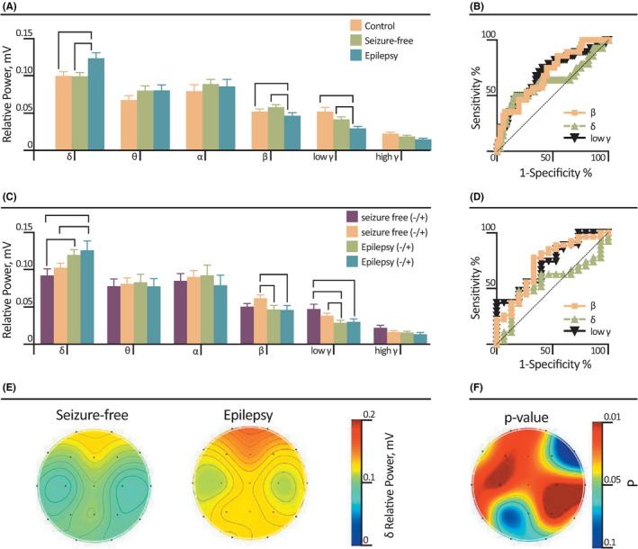
Objective: Management of a patient presenting with a first seizure depends on the risk of additional seizures. In clinical practice, the recurrence risk is estimated by the treating physician using the neurological examination, brain imaging, a thorough history for risk factors, and routine scalp electroencephalogram (EEG) to detect abnormal epileptiform activity. The decision to use antiseizure medication can be challenging when objective findings are missing. There is a need for new biomarkers to better diagnose epilepsy following a first seizure. Recently, an EEG-based novel analytical method was reported to detect paroxysmal slowing in the cortical network of patients with epilepsy. The aim of our study is to test this method's sensitivity and specificity to predict epilepsy following a first seizure.
Methods: We analyzed interictal EEGs of 70 patients admitted to the emergency department of a tertiary referral center after a first seizure. Clinical data from a follow-up period of at least 18 months were available. EEGs of 30 healthy controls were also analyzed and included. For each EEG, we applied an automated algorithm to detect paroxysmal slow wave events (PSWEs).
Results: Of patients presenting with a first seizure, 40% had at least one additional recurring seizure and were diagnosed with epilepsy. Sixty percent did not report additional seizures. A significantly higher occurrence of PSWEs was detected in the first interictal EEG test of those patients who were eventually diagnosed with epilepsy. Conducting the EEG test within 72 h after the first seizure significantly increased the likelihood of detecting PSWEs and the predictive value for epilepsy up to 82%.
Significance: The quantification of PSWEs by an automated algorithm can predict epilepsy and help the neurologist in evaluating a patient with a first seizure.
Keywords: biomarker; epilepsy; first seizure; interictal EEG; new onset seizure; paroxysmal slow wave event.
© 2021 The Authors. Epilepsia published by Wiley Periodicals LLC on behalf of International League Against Epilepsy.
Conflict of interest statement
None of the authors has any conflict of interest to disclose.
Figures


Similar articles
-
Paroxysmal Slow-Wave Events Are Uncommon in Parkinson's Disease.Sensors (Basel). 2023 Jan 13;23(2):918. doi: 10.3390/s23020918. Sensors (Basel). 2023. PMID: 36679715 Free PMC article.
-
Paroxysmal cortical slowing linked to drug-resistant epilepsy.EBioMedicine. 2025 Jun;116:105780. doi: 10.1016/j.ebiom.2025.105780. Epub 2025 May 28. EBioMedicine. 2025. PMID: 40440914 Free PMC article.
-
Quantitative EEG signatures in patients with and without epilepsy development after a first seizure.Epilepsia Open. 2025 Apr;10(2):427-440. doi: 10.1002/epi4.13128. Epub 2025 Mar 4. Epilepsia Open. 2025. PMID: 40040314 Free PMC article.
-
Prognostic significance of interictal epileptiform discharges in newly diagnosed seizure disorders.J Clin Neurophysiol. 2010 Aug;27(4):239-48. doi: 10.1097/WNP.0b013e3181ea4288. J Clin Neurophysiol. 2010. PMID: 20634717 Review.
-
Electroencephalography in Epilepsy Evaluation.Continuum (Minneap Minn). 2019 Apr;25(2):431-453. doi: 10.1212/CON.0000000000000705. Continuum (Minneap Minn). 2019. PMID: 30921017 Review.
Cited by
-
Paroxysmal Cortical Slowing Predicts Posttraumatic Epilepsy After Severe Traumatic Brain Injury.Neurocrit Care. 2025 May 20. doi: 10.1007/s12028-025-02282-5. Online ahead of print. Neurocrit Care. 2025. PMID: 40394356
-
Computer-assisted analysis of routine EEG to identify hidden biomarkers of epilepsy: A systematic review.Comput Struct Biotechnol J. 2023 Dec 10;24:66-86. doi: 10.1016/j.csbj.2023.12.006. eCollection 2024 Dec. Comput Struct Biotechnol J. 2023. PMID: 38204455 Free PMC article. Review.
-
Research progress of epileptic seizure prediction methods based on EEG.Cogn Neurodyn. 2024 Oct;18(5):2731-2750. doi: 10.1007/s11571-024-10109-w. Epub 2024 May 7. Cogn Neurodyn. 2024. PMID: 39555266 Review.
-
Paroxysmal Slow-Wave Events Are Uncommon in Parkinson's Disease.Sensors (Basel). 2023 Jan 13;23(2):918. doi: 10.3390/s23020918. Sensors (Basel). 2023. PMID: 36679715 Free PMC article.
-
Paroxysmal cortical slowing linked to drug-resistant epilepsy.EBioMedicine. 2025 Jun;116:105780. doi: 10.1016/j.ebiom.2025.105780. Epub 2025 May 28. EBioMedicine. 2025. PMID: 40440914 Free PMC article.
References
-
- Jacoby A, Gamble C, Doughty J, Marson A, Chadwick D, Medical Research Council MESS Study Group . Quality of life outcomes of immediate or delayed treatment of early epilepsy and single seizures. Neurology. 2007;10(68):1188–96. - PubMed
-
- Bouma HK, Labos C, Gore GC, Wolfson C, Keezer MR. The diagnostic accuracy of routine electroencephalography after a first unprovoked seizure. Eur J Neurol. 2016;23:455–63. - PubMed
-
- Ikeda A, Takeyama H, Bernard C, Nakatani M, Shimotake A, Daifu M, et al. Active direct current (DC) shifts and “red slow”: two new concepts for seizure mechanisms and identification of the epileptogenic zone. Neurosci Res. 2020;156:95–101. - PubMed
