Review
. 2021 Oct 21:14:767219.
doi: 10.3389/fnmol.2021.767219. eCollection 2021.

Mitochondrial Extracellular Vesicles - Origins and Roles

Affiliations
Review

Mitochondrial Extracellular Vesicles - Origins and Roles

Lydia Amari et al. Front Mol Neurosci. .

Abstract

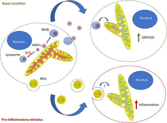
Extracellular vesicles (EVs) have emerged in the last decade as critical cell-to-cell communication devices used to carry nucleic acids and proteins between cells. EV cargo includes plasma membrane and endosomal proteins, but EVs also contain material from other cellular compartments, including mitochondria. Within cells, mitochondria are responsible for a large range of metabolic reactions, but they can also produce damaging levels of reactive oxygen species and induce inflammation when damaged. Consistent with this, recent evidence suggests that EV-mediated transfer of mitochondrial content alters metabolic and inflammatory responses of recipient cells. As EV mitochondrial content is also altered in some pathologies, this could have important implications for their diagnosis and treatment. In this review, we will discuss the nature and roles of mitochondrial EVs, with a special emphasis on the nervous system.

Keywords: extracellular vesicle; inflammation; metabolism; mitochondria; mitochondrial quality control.


Conflict of interest statement

The authors declare that the research was conducted in the absence of any commercial or financial relationships that could be construed as a potential conflict of interest.

Figures

FIGURE 1
FIGURE 1
Model for the roles and mechanisms of secretion of mitochondrial EVs. Under basal conditions, MDVs are required to package mitochondrial components into mitochondrial EVs, which serve to increase OXPHOS in recipient cells. In this setting, oxidized mitochondrial components are not released into EVs but rather sent to lysosomes for degradation through a different type of MDV (Parkin-dependent). On the other hand, pro-inflammatory stimuli like LPS cause the release of free mitochondria or mitochondria packaged within microvesicles that further stimulate inflammation.

References

    1. Al Amir Dache Z., Otandault A., Tanos R., Pastor B., Meddeb R., Sanchez C., et al. (2020). Blood contains circulating cell-free respiratory competent mitochondria. FASEB J. 34 3616–3630. 10.1096/fj.201901917RR - DOI - PubMed
    1. Bernimoulin M., Waters E. K., Foy M., Steele B. M., Sullivan M., Falet H., et al. (2009). Differential stimulation of monocytic cells results in distinct populations of microparticles. J. Thromb. Haemost. 7 1019–1028. 10.1111/j.1538-7836.2009.03434.x - DOI - PMC - PubMed
    1. Boudreau L. H., Duchez A.-C., Cloutier N., Soulet D., Martin N., Bollinger J., et al. (2014). Platelets release mitochondria serving as substrate for bactericidal group IIA-secreted phospholipase A2 to promote inflammation. Blood 124 2173–2183. 10.1182/blood-2014-05-573543 - DOI - PMC - PubMed
    1. Brestoff J. R., Wilen C. B., Moley J. R., Li Y., Zou W., Malvin N. P., et al. (2021). Intercellular mitochondria transfer to macrophages regulates white adipose tissue homeostasis and is impaired in obesity. Cell Metab. 33 270.e8–282.e8. 10.1016/j.cmet.2020.11.008 - DOI - PMC - PubMed
    1. Choong C.-J., Okuno T., Ikenaka K., Baba K., Hayakawa H., Koike M., et al. (2020). Alternative mitochondrial quality control mediated by extracellular release. Autophagy 10.1080/15548627.2020.1848130 [Epub ahead of print]. - DOI - PMC - PubMed