[Preprint]. 2021 Oct 29:2021.10.27.465996.
doi: 10.1101/2021.10.27.465996.

Camel nanobodies broadly neutralize SARS-CoV-2 variants

Affiliations

Camel nanobodies broadly neutralize SARS-CoV-2 variants

Jessica Hong et al. bioRxiv. .

Update in

  • Dromedary camel nanobodies broadly neutralize SARS-CoV-2 variants.
    Hong J, Kwon HJ, Cachau R, Chen CZ, Butay KJ, Duan Z, Li D, Ren H, Liang T, Zhu J, Dandey VP, Martin NP, Esposito D, Ortega-Rodriguez U, Xu M, Borgnia MJ, Xie H, Ho M. Hong J, et al. Proc Natl Acad Sci U S A. 2022 May 3;119(18):e2201433119. doi: 10.1073/pnas.2201433119. Epub 2022 Apr 27. Proc Natl Acad Sci U S A. 2022. PMID: 35476528 Free PMC article.

Abstract

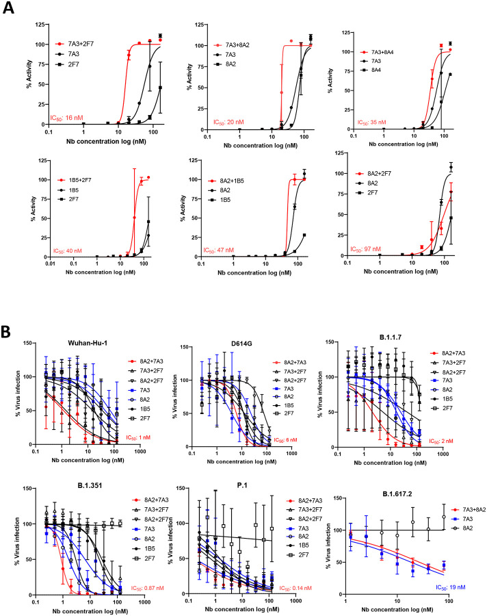
With the emergence of SARS-CoV-2 variants, there is urgent need to develop broadly neutralizing antibodies. Here, we isolate two V H H nanobodies (7A3 and 8A2) from dromedary camels by phage display, which have high affinity for the receptor-binding domain (RBD) and broad neutralization activities against SARS-CoV-2 and its emerging variants. Cryo-EM complex structures reveal that 8A2 binds the RBD in its up mode and 7A3 inhibits receptor binding by uniquely targeting a highly conserved and deeply buried site in the spike regardless of the RBD conformational state. 7A3 at a dose of ≥5 mg/kg efficiently protects K18-hACE2 transgenic mice from the lethal challenge of B.1.351 or B.1.617.2, suggesting that the nanobody has promising therapeutic potentials to curb the COVID-19 surge with emerging SARS-CoV-2 variants.

One-sentence summary: Dromedary camel ( Camelus dromedarius ) V H H phage libraries were built for isolation of the nanobodies that broadly neutralize SARS-CoV-2 variants.


Conflict of interest statement

Competing interests: M.H. and J.H. are inventors on the provisional patent application no. PCT/US2021/056548, “Single domain antibodies targeting SARS coronavirus spike protein and uses thereof.” The authors declare no other competing interests.

Figures

Fig. 1.
Fig. 1.. Isolation of high-affinity camel nanobodies against SARS-CoV-2.
(A) Isolation of camel VHH nanobodies that bind the RBD phage display. (B) Camel VHH nanobodies against the S protein of SARS-CoV-2 or SARS-CoV. (C) Flow cytometry was performed to monitor the cross-reaction of nanobodies to the spike of both SARS-CoV-2 and SARS-CoV on cells. Outline of the experimental workflow for overexpression of SARS-CoV-2-spike or SARS-CoV-spike in the A431 human cell line. Both cell lines were stained with VHH nanobodies or CR3022 as a positive control. (D) Affinity binding (KD) of VHH-hFc antibodies against wild-type SARS-CoV-2 and mutants. The B.1.1.7 variant mutations include HV69–70 deletion, Y144 deletion, N501Y, A570D, D614G, P681H. The B.1.351 variant mutations include K417N, E484K, N501Y, D614G. The P.1. variant includes the E484K mutation. The B.1.617.2 variant includes the T19R, Δ (157–158), L452R, T478K, D614G, P681R, D950N mutations (E) Cross competition assay of each single domain antibody and ACE2 on Octet.
Fig. 2.
Fig. 2.. Nanobodies neutralize SARS-CoV-2 and the variants in pseudovirus assay
(A) Diagrams illustrating pseudovirus assay and VHH-hFc. (B) Camel VHH-hFc proteins inhibit SARS-CoV-2 pseudovirus infectivity to ACE2 expressing human cells by measuring luciferase expression. (C) Pseudovirus particle neutralization assay testing 2-in-1 combination and single nanobodies showing that 7A3+8A2 combination has the best neutralization activity.
Fig. 3.
Fig. 3.. Neutralization of SARS-CoV-2 and its variants in live virus assay
(A) Live virus neutralization assay of nanobodies 7A3, 8A2, 2F7, 1B5, and 8A4 along with their 2-in-1 combination against Wuhan-Hu-1 strain. (B) Live variant virus assay using top three nanobody combinations of 8A2+7A3, 8A2+2F7, and 7A3+2F7 was conducted against SARS-CoV-2 (USA-WA1/2020), D614G variant, B.1.1.7 variant, B.1.351 variant, P.1 variant, and B.1.617.2 variant.
Fig. 4.
Fig. 4.. Protection of the K18-hACE2 mice infected with a lethal dose of B.1.351 or B.1.617.2 variant.
(A) The histology of lung and brain tissues harvested from K18-hACE2 transgenic mice on day 6–7 following infection of live SARS-CoV-2 B.1.351 or B.1.617.2 variant virus. (B) K18-hACE2 mice (n=4/group) were injected intraperitoneally with nanobody 7A3, 8A2 or 2-in-1 cocktail at indicated doses followed by a lethal infection of B.1.351 or B.1.617.2 strain. Mortality and body weight (BW) were monitored for 2-week post infection. (C) Spike-specific IgG titers of K18-hACE2 mice surviving the lethal B.1.351 or B.1.617.2 infection. -: mice died during infection.
Fig. 5.
Fig. 5.. Cryo-EM structure of 7A3 and 8A2 nanobodies with SARS-Cov-2 spike.
(A) The structure models based on the EM images show that 8A2 and 7A3 bind two distinct sites on the RBD. 8A2 directly interferes with the ACE3 binding to the RBD. 7A3 binds a distinct conserved site on the RBD regardless of its conformational mode. 7A3 partially overlaps the ACE2 binding loci when bridging two RBDs. This interaction does not interfere with 8A2 binding, which is only observed in the up conformation. The mutations on the RBD derived from alpha (E484K, S494P, N501Y), beta (K417N, E484K, N501Y), gamma (K417T, E484K, N501Y), and delta (K417N, L452R, and T478K) were indicated. Green spheres, glycans. (B) The unique 7A3 binding pattern. The tip of 7A3 (7A3_A) is deeply buried in the structure of the spike, where the VHH nanobody engages Asp985/B, Glu988/B, Pro987B and Pro384/A in the S2 subunit of the spike protein outside the RBD region. (C) Nanobodies epitopes coverage as determined from the experimental structure using a 5A contact cutoff. Sequences of coronavirus RBD for Bat_RaTG13 (A0A6B9WHD3); Human BJ01 (Q6GYR1); Pangolin (A0A6G6A2Q2); SARS-CoV-2 (P0DTC2) and SARS-CoV-1 (P59594) are presented for comparison purpose. Degree of sequence identity is indicated by background pink hue. 7A3 contact is indicated in blue and 8A2 in orange. Arrows indicate mutation sites of concern (black) and key ACE2 contact residues (green). RBM is indicated by the dotted box. Sequence alignment and colorization was performed using Jalview version 2.11.1. Alignments were performed using ClustallO and colorization by Blosum62.

References

    1. Li Q. et al., Early Transmission Dynamics in Wuhan, China, of Novel Coronavirus-Infected Pneumonia. The New England journal of medicine 382, 1199–1207 (2020). - PMC - PubMed
    1. Zhou F. et al., Clinical course and risk factors for mortality of adult inpatients with COVID-19 in Wuhan, China: a retrospective cohort study. Lancet 395, 1054–1062 (2020). - PMC - PubMed
    1. Vaduganathan M. et al., Renin-Angiotensin-Aldosterone System Inhibitors in Patients with Covid-19. The New England journal of medicine 382, 1653–1659 (2020). - PMC - PubMed
    1. Yan R. et al., Structural basis for the recognition of SARS-CoV-2 by full-length human ACE2. Science 367, 1444–1448 (2020). - PMC - PubMed
    1. Yuan M. et al., A highly conserved cryptic epitope in the receptor-binding domains of SARS-CoV-2 and SARS-CoV. Science, eabb7269 (2020). - PMC - PubMed

Publication types