. 2021 Dec 24;374(6575):1621-1626.
doi: 10.1126/science.abl8506. Epub 2021 Nov 9.

Molecular basis of immune evasion by the Delta and Kappa SARS-CoV-2 variants

Affiliations

Molecular basis of immune evasion by the Delta and Kappa SARS-CoV-2 variants

Matthew McCallum et al. Science. .

Abstract

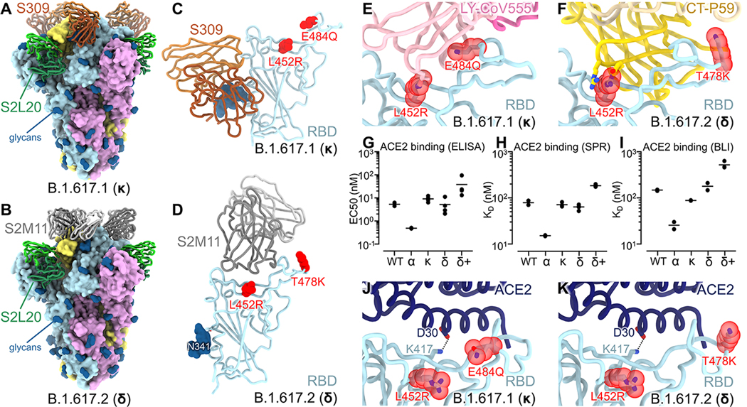
Severe acute respiratory syndrome coronavirus 2 (SARS-CoV-2) transmission leads to the emergence of variants, including the B.1.617.2 (Delta) variant of concern that is causing a new wave of infections and has become globally dominant. We show that these variants dampen the in vitro potency of vaccine-elicited serum neutralizing antibodies and provide a structural framework for describing their immune evasion. Mutations in the B.1.617.1 (Kappa) and Delta spike glycoproteins abrogate recognition by several monoclonal antibodies via alteration of key antigenic sites, including remodeling of the Delta amino-terminal domain. The angiotensin-converting enzyme 2 binding affinities of the Kappa and Delta receptor binding domains are comparable to the Wuhan-Hu-1 isolate, whereas B.1.617.2+ (Delta+) exhibits markedly reduced affinity.


Conflict of interest statement

Competing interests: The remaining authors declare that the research was conducted in the absence of any commercial or financial relationships that could be construed as a potential conflict of interest.

Figures

Figure 1.
Figure 1.. Neutralization of pseudotyped viruses harboring SARS-CoV-2 G614 S, B.1.617.1 S, B.1.617.2 S, or B.1.617.2+ S and incidence of variants.
(A-C) Pairwise connected 50% inhibition concentrations (IC50s) for each individual against each variant for Pfizer/BioNtech BNT162b2 (A), Moderna mRNA-1273 (B), or Janssen Ad26.COV2.S (C) vaccinee sera (Fig. S1 and Table S2). Data are an average of n = 2 replicates and are representative of at least two independent assays with distinct batches of pseudoviruses. Dashed line indicates the limit of detection for the assay. Means (shown as thick black horizontal lines) were compared against G614 by two-way ANOVA (Dunnet’s test); *, p<0.05; ***, p<0.001; ****, p<0.0001. (D-F) Incidence (7-day average) of variants of concern and variants of interest as a proportion of viruses sequenced in the world (D), India (E), and the USA (F) deposited to GISAID (analyzed using outbreak.info) from December 1, 2020 to September 30, 2021. B.1.1.7 (alpha, α), B.1.351 (beta, β), P.1 (gamma, γ), B.1.617.2 (delta, δ) including AY.3-AY.31, B.1.526 (iota, ι), B.1.427/B.1.429 (epsilon, ε), B.1.617.1 (kappa, κ), and B.1.617.2+ (delta+, δ+) including AY.1 and AY.2 are shown in yellow, light purple, cyan, red, blue, green, orange, and dark purple, respectively.
Figure 2.
Figure 2.. CryoEM structures of the SARS-CoV-2 B.1.617.1 and B.1.617.2 S ectodomain trimers and analysis of ACE2 binding.
(A-B) Structure of the B.1.617.1 (A) and B.1.617.2 (B) S trimer (surface rendering) bound to the S2L20 and S309 (A) or S2M11 (B) Fabs (ribbons). SARS-CoV-2 S protomers are colored pink, cyan, and gold, whereas the S2L20 Fab heavy and light chains are colored dark and light green, respectively. The S309 Fab heavy and light chains are colored dark and light orange, respectively (A). The S2M11 Fab heavy and light chains are colored dark and light gray, respectively (B). Only the Fab variable domains are resolved and therefore modeled in the map. N-linked glycans are rendered as dark blue spheres. (C) Zoomed in view of the S309-bound B.1.617.1 RBD with L452R and E484Q shown as red spheres. (D) Zoomed in view of the S2M11-bound B.1.617.2 RBD with L452R and T478K shown as red spheres. (E) Superimposition of the LY-CoV555–bound SARS-CoV-2 RBD structure (PDB 7KMG) on the SARS-CoV-2 B.1.617.1 S cryoEM structure show that L452R would clash with the mAb and E484Q would disrupt electrostatic interactions. (F) Superimposition of the CT-P59–bound SARS-CoV-2 RBD structure (PDB 7CM4) on the SARS-CoV-2 B.1.617.2 S cryoEM structure show that L452R would sterically clash with the mAb. (G) Enzyme-linked immunosorbant assay (ELISA) binding analysis of the SARS-CoV-2 wildtype, B.1.1.7 (α), B.1.617.1 (κ), B.1.617.2 (δ), and B.1.617.2+ (δ+) RBDs to immobilized human ACE2 ectodomain (residues 1–615) shown as 50% effective concentrations (EC50). Data from two biological replicates are shown with 2–4 technical replicates each. (H) Surface plasmon resonance (SPR) binding affinity analysis of the human ACE2 ectodomain (residues 1–615) for immobilized biotinylated wildtype, B.1.1.7 (α), B.1.617.1 (κ), B.1.617.2 (δ), and B.1.617.2+ (δ+) RBDs. Data from two biological replicates are shown with 2–6 technical replicates each. (I) Biolayer interferometry (BLI) binding analysis of the human ACE2 ectodomain (residues 1–615) to immobilized biotinylated SARS-CoV-2 wildtype, B.1.1.7 (α), B.1.617.1 (κ), B.1.617.2 (δ), and B.1.617.2+ (δ+) RBDs. Data from two biological replicates are shown with 1–2 technical replicates each. (J-K) Superimposition of the ACE2-bound SARS-CoV-2 RBD structure (PDB 6VW1) on the SARS-CoV-2 B.1.617.1 (J) and B.1.617.2 (K) S cryoEM structures show that L452R and T478K point away from the interface with ACE2, while K417 contacts D30 from ACE2.
Figure 3.
Figure 3.. Remodeling of the NTD antigenic supersite in the B.1.617.1 and B.1.617.2 S variants.
A-B, Ribbon diagrams of the B.1.617.1 (A) and B.1.617.2 (B) NTDs in the same orientation. Mutated residues are rendered as red spheres and N-linked glycans are shown as dark blue surfaces. Segments with notable structural changes as a consequence of these mutated residues are shown in orange and labeled. C-D, Zoomed-in views of the B.1.617.1 (C) and B.1.617.2 (D) NTD antigenic supersites highlighting incompatibility with recognition by the S2X333 mAb (23) (used here as an example of prototypical NTD neutralizing mAb). N- and C- termini are labeled.
Figure 4.
Figure 4.. S2X303 defines a subclass of site i NTD mAbs cross-reacting with several variants.
(A) Binding of a panel of 11 neutralizing (antigenic site i) and 1 non-neutralizing (S2L20, antigenic site iv) NTD-specific mAbs to recombinant SARS-CoV-2 S variants analyzed by ELISA displayed as a heat map (relative to wildtype Wuhan-Hu-1 binding). (B) Structure of the B.1.617.1 S trimer (surface rendering) bound to the S2X303 Fab fragment (ribbons) shown in two orthogonal orientations. SARS-CoV-2 S protomers are colored pink, cyan, and gold, whereas the S2X303 Fab heavy and light chains are colored dark and light purple, respectively. Only the Fab variable domains are resolved and therefore modeled in the map. N-linked glycans are rendered as dark blue spheres. (C) Ribbon diagram of the S2X303-bound SARS-CoV-2 B.1.617.1 NTD. (D) Zoomed-in view of the S2X303-bound B.1.617.1 NTD with key residues involved in the interface shown as sticks. (E) Structure of the S trimer bound to the S2X303 overlaid with S2X333 and P008–056 antibodies (PDB IDs 7LXW and 7NTC, respectively) shown as a surface rendering. S is colored as in (B); S2X303, S2X333, and P008–056 are shown in purple, orange, and light grey, respectively.

Update of

References

    1. Cele S, Gazy I, Jackson L, Hwa S-H, Tegally H, Lustig G, Giandhari J, Pillay S, Wilkinson E, Naidoo Y, Karim F, Ganga Y, Khan K, Bernstein M, Balazs AB, Gosnell BI, Hanekom W, Moosa M-YS, NGS-SA, COMMIT-KZN Team, Lessells RJ, de Oliveira T, Sigal A, Escape of SARS-CoV-2 501Y.V2 from neutralization by convalescent plasma. Nature (2021), doi: 10.1038/s41586-021-03471-w. - DOI - PMC - PubMed
    1. Tegally H, Wilkinson E, Giovanetti M, Iranzadeh A, Fonseca V, Giandhari J, Doolabh D, Pillay S, San EJ, Msomi N, Mlisana K, von Gottberg A, Walaza S, Allam M, Ismail A, Mohale T, Glass AJ, Engelbrecht S, Van Zyl G, Preiser W, Petruccione F, Sigal A, Hardie D, Marais G, Hsiao M, Korsman S, Davies M-A, Tyers L, Mudau I, York D, Maslo C, Goedhals D, Abrahams S, Laguda-Akingba O, Alisoltani-Dehkordi A, Godzik A, Wibmer CK, Sewell BT, Lourenço J, Alcantara LCJ, Kosakovsky Pond SL, Weaver S, Martin D, Lessells RJ, Bhiman JN, Williamson C, de Oliveira T, Emergence of a SARS-CoV-2 variant of concern with mutations in spike glycoprotein. Nature (2021), doi: 10.1038/s41586-021-03402-9. - DOI - PubMed
    1. Wibmer CK, Ayres F, Hermanus T, Madzivhandila M, Kgagudi P, Oosthuysen B, Lambson BE, de Oliveira T, Vermeulen M, van der Berg K, Rossouw T, Boswell M, Ueckermann V, Meiring S, von Gottberg A, Cohen C, Morris L, Bhiman JN, Moore PL, SARS-CoV-2 501Y.V2 escapes neutralization by South African COVID-19 donor plasma. Nat. Med (2021), doi: 10.1038/s41591-021-01285-x. - DOI - PubMed
    1. Collier DA, De Marco A, Ferreira IATM, Meng B, Datir R, Walls AC, Kemp S SA, Bassi J, Pinto D, Fregni CS, Bianchi S, Tortorici MA, Bowen J, Culap K, Jaconi S, Cameroni E, Snell G, Pizzuto MS, Pellanda AF, Garzoni C, Riva A, Elmer A, Kingston N, Graves B, McCoy LE, Smith KGC, Bradley JR, Temperton N, Lourdes Ceron-Gutierrez L, Barcenas-Morales G, Harvey W, Virgin HW, Lanzavecchia A, Piccoli L, Doffinger R, Wills M, Veesler D, Corti D, Gupta RK, The CITIID-NIHR BioResource COVID-19 Collaboration, The COVID-19 Genomics UK (COG-UK) consortium, Sensitivity of SARS-CoV-2 B.1.1.7 to mRNA vaccine-elicited antibodies. Nature (2021), doi: 10.1038/s41586-021-03412-7. - DOI - PMC - PubMed
    1. Walls AC, Park YJ, Tortorici MA, Wall A, McGuire AT, Veesler D, Structure, Function, and Antigenicity of the SARS-CoV-2 Spike Glycoprotein. Cell. 181, 281–292.e6 (2020). - PMC - PubMed

Publication types

MeSH terms

Supplementary concepts