Review
. 2022 Apr 1;95(1132):20210824.
doi: 10.1259/bjr.20210824. Epub 2021 Nov 9.

Overview of positron emission tomography in functional imaging of the lungs for diffuse lung diseases

Affiliations
Review

Overview of positron emission tomography in functional imaging of the lungs for diffuse lung diseases

Avanti V Gulhane et al. Br J Radiol. .

Abstract

Positron emission tomography (PET) is a quantitative molecular imaging modality increasingly used to study pulmonary disease processes and drug effects on those processes. The wide range of drugs and other entities that can be radiolabeled to study molecularly targeted processes is a major strength of PET, thus providing a noninvasive approach for obtaining molecular phenotyping information. The use of PET to monitor disease progression and treatment outcomes in DLD has been limited in clinical practice, with most of such applications occurring in the context of research investigations under clinical trials. Given the high costs and failure rates for lung drug development efforts, molecular imaging lung biomarkers are needed not only to aid these efforts but also to improve clinical characterization of these diseases beyond canonical anatomic classifications based on computed tomography. The purpose of this review article is to provide an overview of PET applications in characterizing lung disease, focusing on novel tracers that are in clinical development for DLD molecular phenotyping, and briefly address considerations for accurately quantifying lung PET signals.


Figures

Figure 1.
Figure 1.
Increased uptake of 68Ga-DOTANOC in peripheral and subpleural areas on PET/CT (A) in a patient with IPF in areas of interstitial fibrosis and honeycombing on axial HRCT chest images in the same patient (B), representing somatostatin receptor expression in lung fibrosis. Modified and Reprinted from JNM Figure 1. https://doiorg/102967/jnumed110079962. This research was originally published in JNM. Author(s): Valentina Ambrosini, Maurizio Zompatori, Fiorella De Luca, D'Errico Antonia, Vincenzo Allegri, Cristina Nanni, Deborah Malvi, Eva Tonveronachi, Luca Fasano, Mario Fabbri and Stefano Fanti; Title 68Ga-DOTANOC PET/CT Allows Somatostatin Receptor Imaging in Idiopathic Pulmonary Fibrosis: Preliminary Results; J Nucl Med. Year 2010,; vol: 51 (12) pp 1950–1955; © SNMMI.
Figure 2.
Figure 2.
18F-NOS PET/CT parametric images of the distribution volume ratio (DVR), calculated using Logan plot analysis (units mL lung/mL blood, scale shown on the right) were obtained before and after bronchoscopic instillation of endotoxin in right middle lobe. Increased signal in areas of segmental neutrophilic inflammation in the right middle lobe correlate with increased infiltrate on CT (white volumes of interest). Modified and Reprinted from JNM Figure 3. https://doi.org/10.2967/jnumed.114.146381 This research was originally published in JNM. Author(s): Howard J. Huang, Warren Isakow, Derek E. Byers, Jacquelyn T. Engle, Elizabeth A. Griffin, Debra Kemp, Steven L. Brody, Robert J. Gropler, J. Philip Miller, Wenhua Chu, Dong Zhou, Richard A. Pierce, Mario Castro, Robert H. Mach and Delphine L. Chen; Title Imaging Pulmonary Inducible Nitric Oxide Synthase Expression with PET; J Nucl Med. Year 2015; vol: 56 (1) pp 76–81; © 2015 by the Society of Nuclear Medicine and Molecular Imaging, Inc.
Figure 3.
Figure 3.
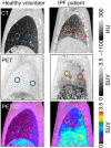
64Cu-DOTA-ECL1i PET/CT to determine binding to macrophage CCR2 receptors in lungs of a healthy volunteer (left panel) and a patient with idiopathic pulmonary fibrosis (IPF, right panel). Blue regions of interest (ROI) denote anterior and posterior regions in healthy lungs for comparison. The orange ROI denotes an area with CT-evident fibrosis, while the yellow ROI denotes an area without CT evidence of fibrosis. Red arrowheads point to subpleural areas of fibrosis and increased uptake. PET only images (middle row) and PET and CT fused images (bottom row) are displayed using the same standardized uptake value (SUV) scale. Increased uptake was seen with increased lung fibrosis in all IPF patients. Adapted with permission of the American Thoracic Society. Copyright © 2021 American Thoracic Society*. All rights reserved. Cite: Brody SL, Gunsten SP, Luehmann HP, Sultan DH, Hoelscher M, Heo GS, Pan J, Koenitzer JR, Lee EC, Huang T, Mpoy C, Guo S, Laforest R, Salter A, Russell TD, Shifren A, Combadiere C, Lavine KJ, Kreisel D, Humphreys BD, Rogers BE, Gierada DS, Byers DE, Gropler RJ, Chen DL, Atkinson JJ, Liu Y. Chemokine Receptor 2-targeted Molecular Imaging in Pulmonary Fibrosis. A Clinical Trial.Am J Respir Crit Care Med. 2021 Jan 1;203(1):78–89. doi: 10.1164/rccm.202004–1132OC. PMID: 32673071; PMCID: PMC7781144. The American Journal of Respiratory and Critical Care Medicine is an official journal of the American Thoracic Society. Readers are encouraged to read the entire article for the correct context at [https://www.atsjournals.org/doi/abs/10.1164/rccm.202004–1132OC]. The authors, editors, and The American Thoracic Society are not responsible for errors or omissions in adaptations. *Now titled Annals of the American Thoracic Society.
Figure 4.
Figure 4.
PET imaging with the cathepsin-targeted tracer 68Ga-BMV101 showing significantly higher uptake in lung areas with increased fibrosis in patients with IPF when compared to normal controls or patients with unclassified pulmonary fibrosis. Modified and reprinted from Withana NP, Ma X, McGuire HM, Verdoes M, van der Linden WA, Ofori LO, Zhang R, Li H, Sanman LE, Wei K, Yao S, Wu P, Li F, Huang H, Xu Z, Wolters PJ, Rosen GD, Collard HR, Zhu Z, Cheng Z, Bogyo M. Non-invasive Imaging of Idiopathic Pulmonary Fibrosis Using Cathepsin Protease Probes. Sci Rep. 2016 Jan 22;6:1,9755. Figure 6. doi: 10.1038/srep19755. PMID: 26797565; PMCID: PMC4726431. This article is distributed under the terms of the Creative Commons Attribution 4.0 International License (http://creativecommons.org/licenses/by/4.0/), which permits unrestricted use, distribution, and reproduction in any medium.
Figure 5.
Figure 5.
PET imaging with the αvβ6 integrin-targeted tracer 18F-FB-A20FMDV2 in a patient with IPF (A,B) and healthy volunteer (C). Uptake in the IPF patient shows significantly higher 18F-FB-A20FMDV2 uptake in fibrotic areas compared to healthy lungs, suggesting integrin αvβ6 expression related to TGF-β related processes in areas of fibrosis. (Reprinted with modification). Modified and reprinted from Lukey PT, Coello C, Gunn R, Parker C, Wilson FJ, Saleem A, Garman N, Costa M, Kendrick S, Onega M, Kang'ombe AR, Listanco A, Davies J, Ramada-Magalhaes J, Moz S, Fahy WA, Maher TM, Jenkins G, Passchier J, Marshall RP. Clinical quantification of the integrin αvβ6 by [18F]FB-A20FMDV2 positron emission tomography in healthy and fibrotic human lung (PETAL Study). Eur J Nucl Med Mol Imaging. 2020 Apr;47(4):967–979. doi: 10.1007/s00259-019-04586-z. Epub 2019 Dec 9. Figure 1. PMID: 31814068; PMCID: PMC7075837. This article is distributed under the terms of the Creative Commons Attribution 4.0 International License (http://creativecommons.org/licenses/by/4.0/), which permits unrestricted use, distribution, and reproduction in any medium.
Figure 6.
Figure 6.
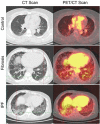
PET and CT images obtained with the type I collagen-targeted tracer 68Ga-CBP8. Minimal uptake is noted in the lungs on representative PET images from a healthy volunteer (A). In contrast, heterogeneously increased uptake is noted throughout the lungs on representative PET images from a patient with IPF (B), with more intense uptake (red arrows) in areas of CT-evident fibrosis (C). Lower-level uptake was also noted in areas without CT evidence of fibrosis (white arrows), suggesting that this approach can identify early fibrotic changes in the lungs. Adapted with permission of the American Thoracic Society. Copyright © 2021 American Thoracic Society*. All rights reserved. Cite: Montesi SB, Izquierdo-Garcia D, Desogere P, Abston E, Liang LL, Digumarthy S, et al. Type I Collagen–targeted Positron Emission Tomography Imaging in Idiopathic Pulmonary Fibrosis: First-in-Human Studies Am J Respir Crit Care Med. 2019 Jul 15; 200(2): 258–261. Figure 2. doi.org/10.1164/rccm.201903-0503LE . PMID: 31161770; PMCID: PMC6635789, Published online 2019 Jul 15 The American Journal of Respiratory and Critical Care Medicine is an official journal of the American Thoracic Society. Readers are encouraged to read the entire article for the correct context at [https://www.atsjournals.org/doi/abs/10.1164/rccm.202004–1132OC]. The authors, editors, and The American Thoracic Society are not responsible for errors or omissions in adaptations. *Now titled Annals of the American Thoracic Society.

Similar articles

Cited by

References

    1. Schuster DP. The evaluation of lung function with PET. Semin Nucl Med 1998; 28: 341–51. doi: 10.1016/s0001-2998(98)80037-8 - DOI - PubMed
    1. Chen DL, Ballout S, Chen L, Cheriyan J, Choudhury G, Denis-Bacelar AM, et al. . Consensus Recommendations on the Use of 18F-FDG PET/CT in Lung Disease. J Nucl Med 2020; 61: 1701–7. doi: 10.2967/jnumed.120.244780 - DOI - PMC - PubMed
    1. Chen DL, Cheriyan J, Chilvers ER, Choudhury G, Coello C, Connell M, et al. . Quantification of lung PET images: challenges and opportunities. J Nucl Med 2017; 58: 201–7. doi: 10.2967/jnumed.116.184796 - DOI - PMC - PubMed
    1. Zhou Z, Kozlowski J, Goodrich AL, Markman N, Chen DL, Schuster DP. Molecular imaging of lung glucose uptake after endotoxin in mice. Am J Physiol Lung Cell Mol Physiol 2005; 289: L760–8. doi: 10.1152/ajplung.00146.2005 - DOI - PubMed
    1. Saha D, Takahashi K, de Prost N, Winkler T, Pinilla-Vera M, Baron RM, et al. . Micro-autoradiographic assessment of cell types contributing to 2-deoxy-2-[(18)F]fluoro-D-glucose uptake during ventilator-induced and endotoxemic lung injury. Mol Imaging Biol 2013; 15: 19–27. doi: 10.1007/s11307-012-0575-x - DOI - PMC - PubMed

Substances