. 2021 Nov 9;19(11):e3001442.
doi: 10.1371/journal.pbio.3001442. eCollection 2021 Nov.

Diversity, taxonomy, and evolution of archaeal viruses of the class Caudoviricetes

Affiliations

Diversity, taxonomy, and evolution of archaeal viruses of the class Caudoviricetes

Ying Liu et al. PLoS Biol. .

Abstract

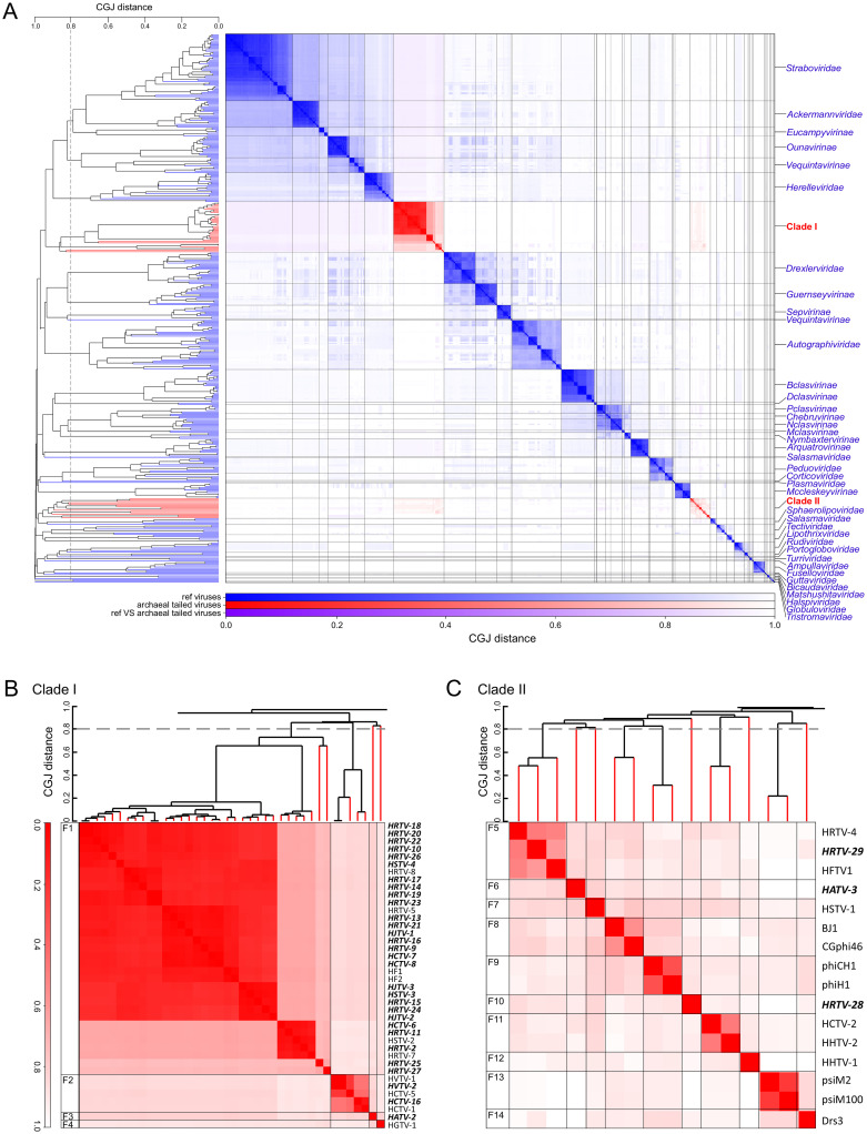
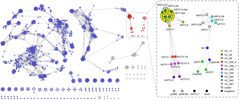
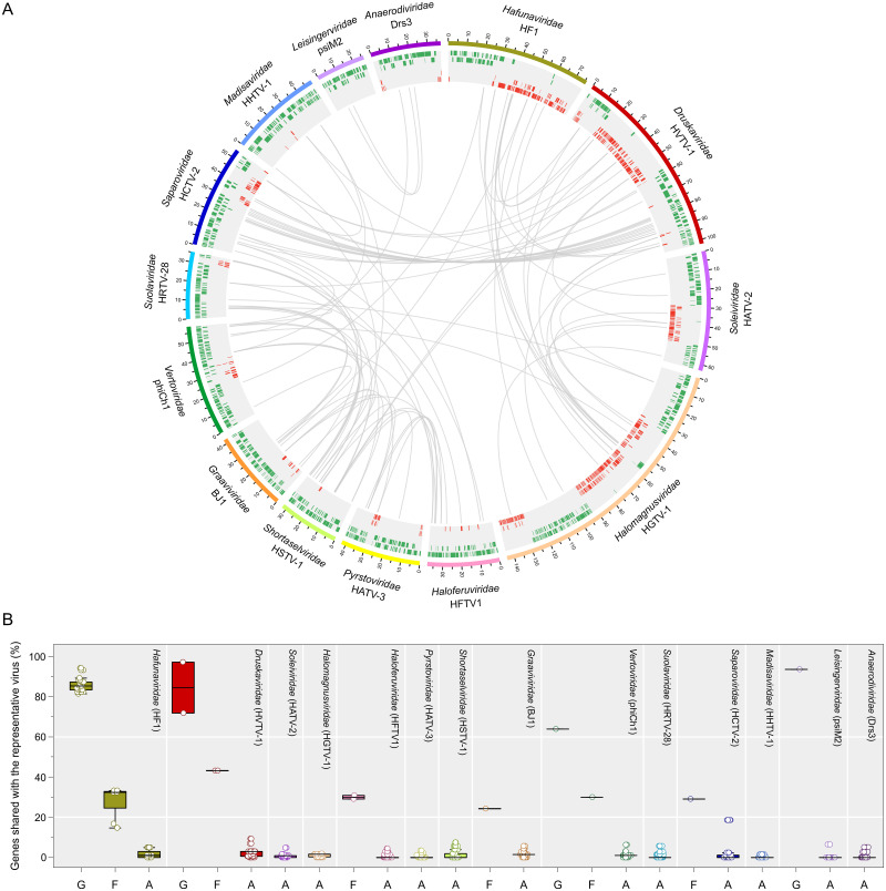
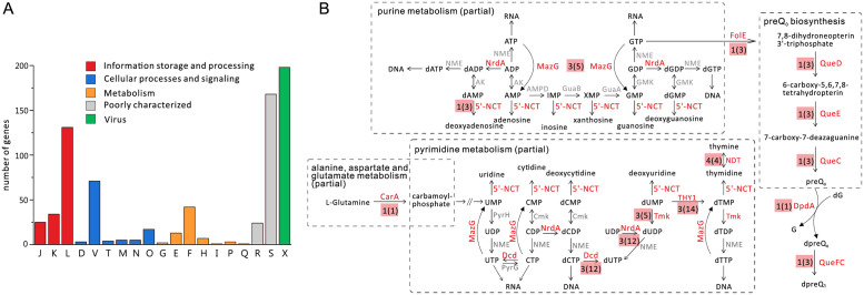
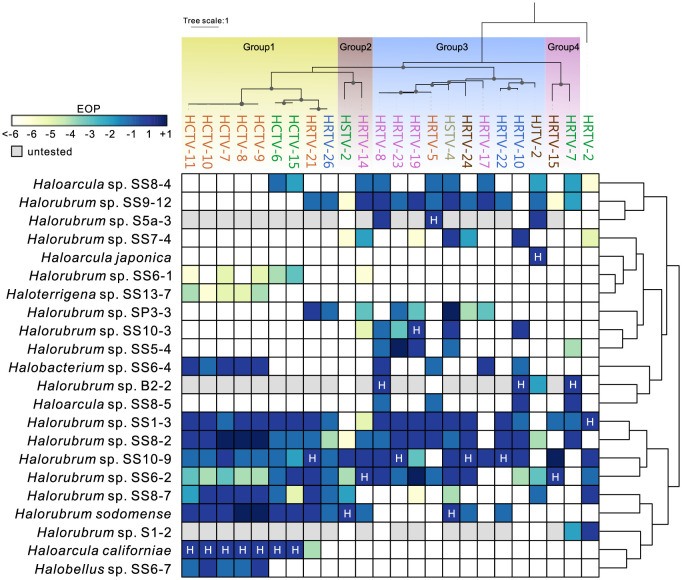
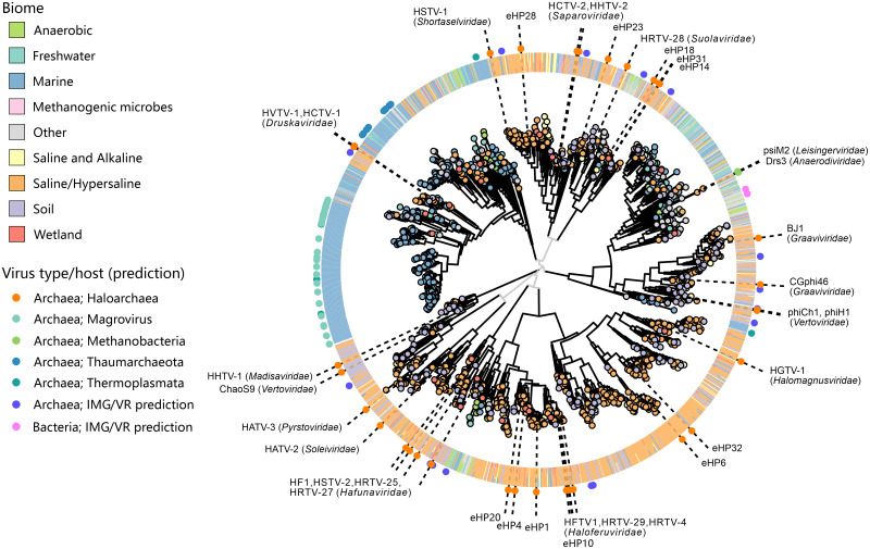
The archaeal tailed viruses (arTV), evolutionarily related to tailed double-stranded DNA (dsDNA) bacteriophages of the class Caudoviricetes, represent the most common isolates infecting halophilic archaea. Only a handful of these viruses have been genomically characterized, limiting our appreciation of their ecological impacts and evolution. Here, we present 37 new genomes of haloarchaeal tailed virus isolates, more than doubling the current number of sequenced arTVs. Analysis of all 63 available complete genomes of arTVs, which we propose to classify into 14 new families and 3 orders, suggests ancient divergence of archaeal and bacterial tailed viruses and points to an extensive sharing of genes involved in DNA metabolism and counterdefense mechanisms, illuminating common strategies of virus-host interactions with tailed bacteriophages. Coupling of the comparative genomics with the host range analysis on a broad panel of haloarchaeal species uncovered 4 distinct groups of viral tail fiber adhesins controlling the host range expansion. The survey of metagenomes using viral hallmark genes suggests that the global architecture of the arTV community is shaped through recurrent transfers between different biomes, including hypersaline, marine, and anoxic environments.


Conflict of interest statement

The authors have declared that no competing interests exist.

Figures

Fig 1
Fig 1. Genome relationships between prokaryotic dsDNA viruses.
(A) Heat map and dendrogram of CGJ distances for classified bacteriophages and archaeal dsDNA viruses. Branches and clusters corresponding to arTVs are shown in red, whereas those of other viruses are in blue. (B) Zoom in on the arTV Clade I. (C) Zoom in on the arTV Clade II. Viruses sequenced in this study are highlighted in bold. CGJ distance of 0.8, chosen as a family-level threshold, is indicated with a broken line, with the family-level groups (F1 to F14) indicated on the left of the heat map. See S1 Data for the underlying dendrogram. arTV, archaeal tailed virus; CGJ, composite generalized Jaccard; dsDNA, double-stranded DNA.
Fig 2
Fig 2. The network-based analysis of PCs shared among arTVs and the prokaryotic dsDNA viruses.
The nodes represent viral genomes, and the edges represent the strength of connectivity between each genome based on shared PCs. Nodes representing genomes of arTVs are in red and other archaeal viruses are in pink, whereas those representing genomes of tailed bacteriophages are in blue and other bacteriophages are in light blue (left panel). The VCs of arTVs are enlarged and labeled in the right panel. See S2 Data for complete VCs and PCs generated by vConTACT v2.0. arTV, archaeal tailed virus; dsDNA, double-stranded DNA; PC, protein cluster; VC, viral cluster.
Fig 3
Fig 3. Overview of homologous proteins shared by arTVs.
(A) The circus plot displays gene content similarity between representative members of each proposed family. Viral genomes from different families are indicated with distinct colors. The genomic coordinates (kbp) are indicated on genome maps. Putative ORFs distributed on 5 tracks (to avoid overlapping) are represented by green and red tiles on the forward and reverse strands, respectively. Homologous proteins are linked by gray lines. The representative viruses and the proposed virus family names are indicated. (B) The box plot shows the percentage of genes shared by a representative virus from each family with other members of the same genus (G) and family (F) as well as with arTVs from other families (A). Each box represents the middle 50th percentile of the data set and is derived using the lower and upper quartile values. The median value is displayed by a horizontal line. Whiskers represent the maximum and minimum values with the range of 1.5 IQR. Each virus is represented by a dot. In the circus and box plots analyses, proteins with over 30% amino acid sequence identity and E-value < 1 × 10⁻²⁵ in our arTV database are considered to be homologous. Data underlying this figure can be found in S2 Data. arTV, archaeal tailed virus.
Fig 4
Fig 4. Classification of genes encoded by arTVs.
(A) Classification of arTV genes into arCOG functional categories. The homologous gene shared by viruses in the same species is counted as one. The letters represent the following: J, translation, ribosomal structure and biogenesis; K, transcription; L, replication, recombination and repair; D, cell cycle control, cell division, chromosome partitioning; V, defense mechanisms; T, signal transduction mechanisms; M, cell wall/membrane/envelope biogenesis; N, cell motility; O, posttranslational modification, protein turnover, chaperons; G, carbohydrate transport and metabolism; E, amino acid transport and metabolism; F, nucleotide transport and metabolism; H, coenzyme transport and metabolism; I, lipid transport and metabolism; P, inorganic ion transport and metabolism; Q, secondary metabolites biosynthesis, transport and catabolism; R, general function prediction only; S, function unknown; X, virus related. (B) Schematic showing nucleotide-related (partial) metabolic pathways, with a particular enzyme indicated for each reaction. Enzymes that are found encoded by arTVs are in red, otherwise in gray. The number of virus families that harbor the enzyme is indicated next to the enzyme name, with the number of virus species shown in parentheses. NrdA: RnR, MazG: NTP pyrophosphatase, 5′-NCT: 5′-deoxyribonucleotidase, CarA: CP synthase small subunit, Dcd: dCTP deaminase, Tmk: thymidylate kinase, THY1: thymidylate synthase thyX, NDT: nucleoside deoxyribosyltransferase, FolE: GTP cyclohydrolase I, QueC: queuosine biosynthesis protein, QueD: 6-pyruvoyl tetrahydropterin synthase, QueE: 7-carboxy-7-deazaguanine synthase, DpdA: paralog of queuine tRNA-ribosyltransferase, QueFC: NADPH-dependent 7-cyano-7deazaguanine reductase. See S3 and S5 Tables for individual genes classification. arCOG, archaeal clusters of orthologous gene; arTV, archaeal tailed virus; CP, carbamoylphosphate; RnR, ribonucleotide diphosphate reductase.
Fig 5
Fig 5. Host ranges of hafunaviruses.
The heat map shows the EOP of hafunavirus isolates on different haloarchaeal strains. The EOP of the virus on its original isolation host has been set to 1 and marked with H. Number 1 is equal to the EOP on the original host. Numbers −1, −2,… refer to 10−1, 10−2,… and +1 refers to 101 when compared to the EOP on the original host, as shown in S8 Table. The isolation host for HRTV-26 is Halorubrum sp. SS13-13, which occasionally did not form a lawn and was not included in this analysis. The titer of HRTV-26 on Halorubrum sp. SS13-13 was 2 × 10⁸ plaque-forming units per milliliter, which was considered here as 1 and used for comparison. The hosts were clustered based on the similarity of EOPs of the tested viruses, as represented by the dendrogram on the right side. The upper panel shows the maximum likelihood phylogenetic tree of the tail fiber adhesin proteins encoded by the analyzed viruses. The adhesin of HRTV-2, the amino acid sequence of which is markedly divergent from those of other hafunaviruses, is set as the outgroup. The 4 distinct groups of adhesins are displayed with colored blocks. Bootstrap values greater than 90% are indicated in the nodes by dots. Virus names are colored according to the species to which they belong. EOP, efficiency of plating.
Fig 6
Fig 6. Phylogenetic analysis of the MCPs encoded by arTVs.
The homologs retrieved from metagenomes were clustered at 0.9 protein sequence identity and the representative of each cluster is indicated as a circle at the tip of the branch with the color reflecting the corresponding biome (see the legend on the left). The ring outside of the tree consists of tiles indicating the biome compositions from the corresponding protein cluster. The dot in the outermost ring indicates the (predicted) virus type in the corresponding branch and is colored based on the host predicted. The names of the arTV isolates of haloarchaea and methanogens as well as the fosmids generated from a saltern of Santa Pola in Spain [75] are shown on the tree. The virus family names are indicated next to the corresponding arTV isolates. The associated data on metagenomic contigs from the IMG/M database can be found in S2 Data. arTV, archaeal tailed virus; IMG/M, Integrated Microbial Genomes and Microbiomes; MCP, major capsid protein.
Fig 7
Fig 7. Structural comparison of the arTV MCPs.
(A) Structural models of arTV MCPs shown using ribbon representation and colored using rainbow scheme from the N-terminus (blue) to C-terminus (red). The models are shown next to the corresponding tips of the cladogram based on the pairwise structural similarity (Z) scores. (B) All-against-all comparison of the arTV MCP structural models shown as a matrix of pairwise Z scores generated using DALI [93]. The underlying data can be found in S2 Data. arTV, archaeal tailed virus; MagroA/B, group A and B Magroviruses; MCP, major capsid protein; Thaum, a virus of marine Thaumarchaeota; Thermopl, a virus of marine Thermoplasmata.

References

    1. Koonin EV, Dolja VV, Krupovic M, Varsani A, Wolf YI, Yutin N, et al.. Global Organization and Proposed Megataxonomy of the Virus World. Microbiol Mol Biol Rev. 2020;84(2):e00061–19. Epub 2020/03/07. doi: 10.1128/MMBR.00061-19 . - DOI - PMC - PubMed
    1. Dion MB, Oechslin F, Moineau S. Phage diversity, genomics and phylogeny. Nat Rev Microbiol 2020;18(3):125–38. Epub 2020/02/06. doi: 10.1038/s41579-019-0311-5 ; - DOI - PubMed
    1. Krupovic M, Dolja VV, Koonin EV. The LUCA and its complex virome. Nat Rev Microbiol 2020;18(11):661–70. Epub 2020/07/16. doi: 10.1038/s41579-020-0408-x ; - DOI - PubMed
    1. Dedeo CL, Cingolani G, Teschke CM. Portal Protein: The Orchestrator of Capsid Assembly for the dsDNA Tailed Bacteriophages and Herpesviruses. Annu Rev Virol. 2019;6(1):141–60. Epub 2019/07/25. doi: 10.1146/annurev-virology-092818-015819 . - DOI - PMC - PubMed
    1. Nobrega FL, Vlot M, de Jonge PA, Dreesens LL, Beaumont HJE, Lavigne R, et al.. Targeting mechanisms of tailed bacteriophages. Nat Rev Microbiol 2018;16(12):760–73. Epub 2018/08/15. doi: 10.1038/s41579-018-0070-8 - DOI - PubMed

Publication types