Review
. 2021 Nov;11(11):210194.
doi: 10.1098/rsob.210194. Epub 2021 Nov 10.

Maintenance and loss of endocytic organelle integrity: mechanisms and implications for antigen cross-presentation

Affiliations
Review

Maintenance and loss of endocytic organelle integrity: mechanisms and implications for antigen cross-presentation

Eleanor Childs et al. Open Biol. 2021 Nov.

Abstract

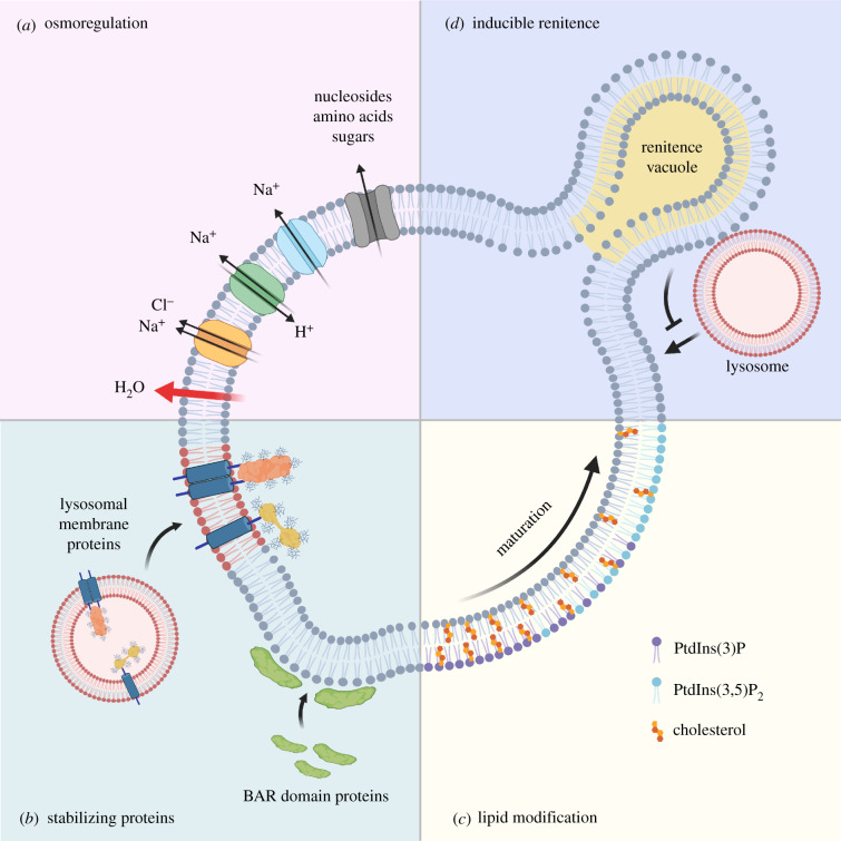
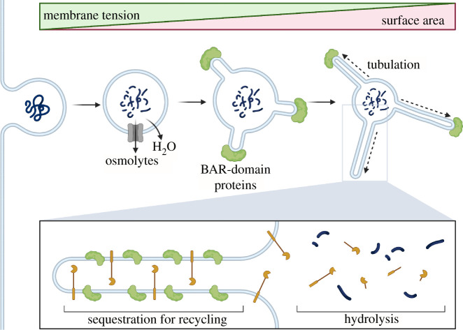
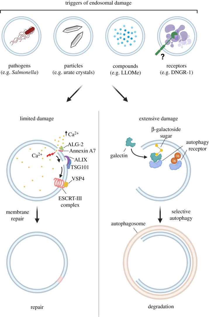
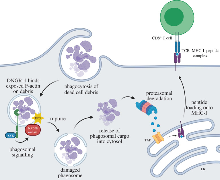
The membranes of endosomes, phagosomes and macropinosomes can become damaged by the physical properties of internalized cargo, by active pathogenic invasion or by cellular processes, including endocytic maturation. Loss of membrane integrity is often deleterious and is, therefore, prevented by mitigation and repair mechanisms. However, it can occasionally be beneficial and actively induced by cells. Here, we summarize the mechanisms by which cells, in particular phagocytes, try to prevent membrane damage and how, when this fails, they repair or destroy damaged endocytic organelles. We also detail how one type of phagocyte, the dendritic cell, can deliberately trigger localized damage to endocytic organelles to allow for major histocompatibility complex class I presentation of exogenous antigens and initiation of CD8+ T-cell responses to viruses and tumours. Our review highlights mechanisms for the regulation of endocytic organelle membrane integrity at the intersection of cell biology and immunology that could be co-opted for improving vaccination and intracellular drug delivery.

Keywords: cross-presentation; endosomal escape; endosome; membrane damage; membrane repair; phagosome.


Conflict of interest statement

C.R.S. is a founder and stockholder of Adendra Therapeutics and owns stock options and/or is a paid consultant for Adendra Therapeutics, Bicara Therapeutics, Montis Biosciences, Oncurious NV, Bicycle Therapeutics and Sosei Heptares. C.R.S. holds a professorship at Imperial College London and honorary professorships at University College London and King's College London. None of these activities is related to this work.

Figures

Figure 1.
Figure 1.
Maintenance of endocytic organelle integrity. (a) Following internalization of cargo, osmotically induced tension is regulated by solute carriers and transporters that move monovalent and divalent cations across endocytic organelle membranes. This results in the efflux of water and a subsequent reduction in membrane tension. (b) The reduction in volume promotes recruitment of curvature-sensitive BAR domain-containing proteins that stabilize curved membranes, while fusion with lysosomes brings in highly glycosylated transmembrane proteins that help shield the membrane from the luminal environment. (c) Modification of lipids regulates organelle trafficking but also facilitates osmoregulation as in the case of phosphatidylinositol 3,5-bisphosphate [PtdIns(3,5)P2], which promotes efflux of monovalent ions through lipid-gated channels. (d) In macrophages, renitence vacuoles (RVs) are associated with phagosomes and are thought to safeguard against direct fusion between lysosomes and damaged phagosomes. Created with Biorender.com.
Figure 2.
Figure 2.
Membrane tubulation induced by BAR domain-containing proteins. Tubulation dependent on BAR superfamily proteins temporarily increases the surface area of limiting membranes to further facilitate osmolyte efflux. It also spatially segregates cargo for recycling away from hydrolytic enzymes. For example, receptors internalized on a macropinosome membrane can be efficiently recycled to the cell surface by tubulation. Created with Biorender.com.
Figure 3.
Figure 3.
Repair and removal of damaged endocytic organelles. Internalization of some agents can inflict damage to endocytic organelle membranes, leading to rapid Ca2+ efflux. Elevated cytosolic Ca2+ is likely sensed by ALG-2/PDCD6, which recruits components of the ESCRT machinery such as ALIX and TSG101, culminating in recruitment of ESCRT-III (e.g. CHMP4B, VPS4) to seal the damaged membrane. In cases of more extensive membrane damage, galectins (-3/-8) bind exposed β-galactoside residues on luminal proteins and provide a recruitment signal for cargo receptors such as NDP52. Cargo receptors can also recognize ubiquitin chains on the damaged membrane. These receptors trigger the formation of LC3-containing autophagic membranes, leading to removal of the damaged organelle via selective autophagy. Created with Biorender.com.
Figure 4.
Figure 4.
DNGR-1 signals to potentiate ROS-dependent rupture of endocytic organelles and favour P2C and XP of dead cell-associated antigens. In dendritic cells expressing DNGR-1, recognition of F-actin exposed by dead cell cargo within endocytic organelles induces DNGR-1 signalling through SYK, leading to NADPH oxidase activation. ROS produced by the NADPH oxidase damages organelles to increase the probability of rupture and release of cargo into the cytoplasm. There, dead cell-associated antigens are degraded by the proteasome machinery and resulting peptides trafficked into the endoplasmic reticulum (ER) via the TAP transporter for loading onto MHC-I molecules. Peptide–MHC-I complexes are then transported to the membrane and presented to CD8+ T cells. Created with Biorender.com.

References

    1. Tauber AI. 2003. Metchnikoff and the phagocytosis theory. Nat. Rev. Mol. Cell Biol. 4, 897-901. (10.1038/nrm1244) - DOI - PubMed
    1. Lewis WH. 1937. Pinocytosis by malignant cells. Am. J. Cancer Res. 29, 666-679. (10.1158/ajc.1937.510) - DOI
    1. Schmid SL, Sorkin A, Zerial M. 2014. Endocytosis: past, present, and future. Cold Spring Harb. Perspect. Biol. 6, a022509. (10.1101/cshperspect.a022509) - DOI - PMC - PubMed
    1. Levin R, Grinstein S, Canton J. 2016. The life cycle of phagosomes: formation, maturation, and resolution. Immunol. Rev. 273, 156-179. (10.1111/imr.12439) - DOI - PubMed
    1. Freeman SA, Grinstein S. 2018. Resolution of macropinosomes, phagosomes and autolysosomes: osmotically driven shrinkage enables tubulation and vesiculation. Traffic 19, 965-974. (10.1111/tra.12614) - DOI - PubMed

Publication types

Substances