. 2022 Jan;227(1):145-158.
doi: 10.1007/s00429-021-02395-5. Epub 2021 Nov 10.

Post-injury ventricular enlargement associates with iron in choroid plexus but not with seizure susceptibility nor lesion atrophy-6-month MRI follow-up after experimental traumatic brain injury

Affiliations

Post-injury ventricular enlargement associates with iron in choroid plexus but not with seizure susceptibility nor lesion atrophy-6-month MRI follow-up after experimental traumatic brain injury

Amna Yasmin et al. Brain Struct Funct. 2022 Jan.

Abstract

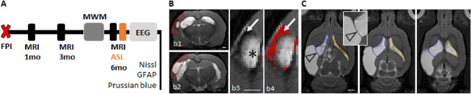
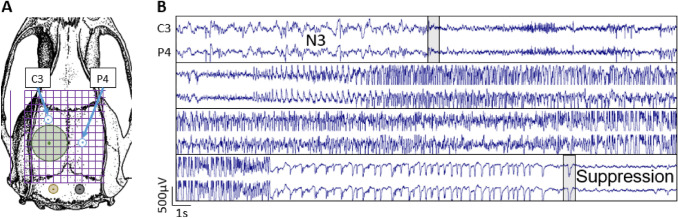
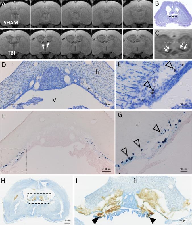
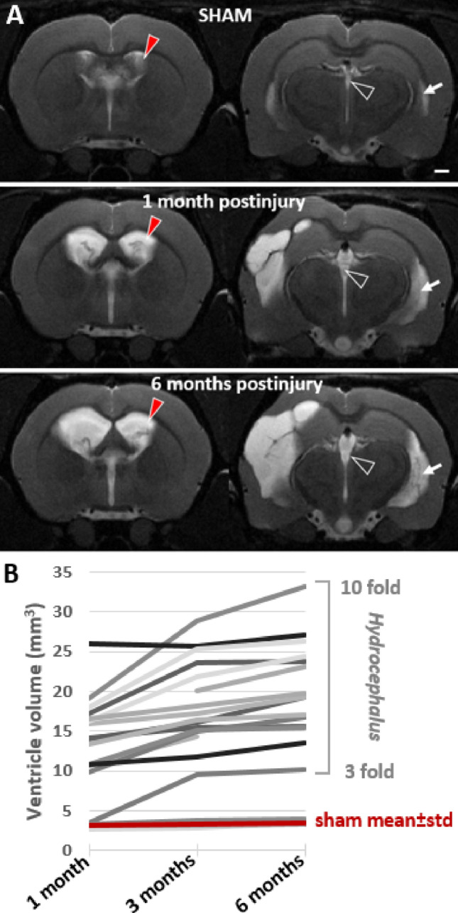
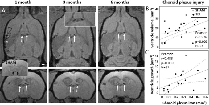
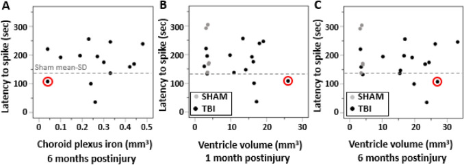
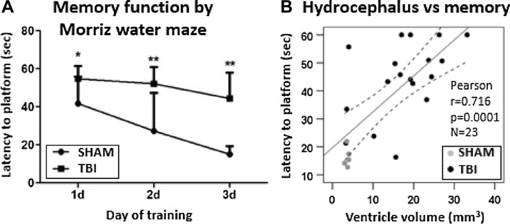
Ventricular enlargement is one long-term consequence of a traumatic brain injury, and a risk factor for memory disorders and epilepsy. One underlying mechanisms of the chronic ventricular enlargement is disturbed cerebrospinal-fluid secretion or absorption by choroid plexus. We set out to characterize the different aspects of ventricular enlargement in lateral fluid percussion injury (FPI) rat model by magnetic resonance imaging (MRI) and discovered choroid plexus injury in rats that later developed hydrocephalus. We followed the brain pathology progression for 6 months and studied how the ventricular growth was associated with the choroid plexus injury, cortical lesion expansion, hemorrhagic load or blood perfusion deficits. We correlated MRI findings with the seizure susceptibility in pentylenetetrazol challenge and memory function in Morris water-maze. Choroid plexus injury was validated by ferric iron (Prussian blue) and cytoarchitecture (Nissl) stainings. We discovered choroid plexus injury that accumulates iron in 90% of FPI rats by MRI. The amount of the choroid plexus iron remained unaltered 1-, 3- and 6-month post-injury. During this time, the ventricles kept on growing bilaterally. Ventricular growth did not depend on the cortical lesion severity or the cortical hemorrhagic load suggesting a separate pathology. Instead, the results indicate choroidal injury as one driver of the post-traumatic hydrocephalus, since the higher the choroid plexus iron load the larger were the ventricles at 6 months. The ventricle size or the choroid plexus iron load did not associate with seizure susceptibility. Cortical hypoperfusion and memory deficits were worse in rats with greater ventricular growth.

Keywords: Brain–CSF barrier; Epileptogenesis; Heme; Idiopathic normal pressure hydrocephalus; Ventriculomegaly.


Conflict of interest statement

The authors declare that the research was conducted in the absence of any commercial or financial relationships that could be construed as a potential conflict of interest.

Figures

Fig. 1
Fig. 1
Study design. A Rats were imaged 1, 3 and 6 months after lateral fluid-percussion injury (FPI). Structural MRI was acquired at each timepoint. Blood perfusion was assessed during the 6 months scan by arterial spin labeling (ASL, described in Supplementary data). Spatial memory was tested at 5 months post-injury by the Morris water-maze (MWM). The pentylenetetrazole seizure-susceptibility test was performed at the end of 4 weeks continuous video-EEG recording. Histology included Nissl (lesion location and ependymal structure) and Prussian blue (ferritic iron) stainings. B Cortical lesion volume and cortical hemorrhagic load were quantified from T2-weighted (T2w) coronal images within a region of interest (ROI) matching with the shape of the intact cortex (red outline in panels b1–b2). Higher magnification images of panel b1 (panels b3 and b4) show the lesion. Both the hyperintense (asterisk in panel b3) and hemorrhagic (arrow in panels b3 and b4) portions were measured and the sum was considered to represent the cortical lesion. Pixels identified to present the hemorrhagic component of the cortical lesion, based on the prior thresholding analysis, are shown in red in panel b4. C Ipsilateral (blue outline) and contralateral (orange outline) ventricle volumes were manually outlined in horizontal T2w images. The insert shows the membrane separating the CSF cavity related to the cortical lesion from the ipsilateral ventricle (arrowhead). Scale bars equal 1 mm in panels b1 and b3, 2 mm in panel C
Fig. 2
Fig. 2
Electroencephalography recordings. A Location of the craniectomy (Green circle 5 mm) and electrodes [two recording electrodes (C3 and P4), ground, and reference]. B Representative example of a generalized seizure that started in the transition between N3 and REM sleep. Beginning and the end of the seizure are indicated by the grey bars
Fig. 3
Fig. 3
T1/T2* mixed-contrast FISP MRI detects post-traumatic iron in the choroid plexus. A Five consecutive 175 µm thick coronal MRI slices (centered at − 1.00 mm from bregma) of the sham-operated (top row) and TBI animal (bottom row) at 6 months post-operation. Note black signal-void areas (white arrows) in the TBI rat which are related to the presence of iron deposits in the choroid plexus. B Thionin-stained section of the same animal sectioned at the corresponding coronal level (7 months post-TBI). C Magnification of the MRI showing the choroidal iron (white arrows). Dashed box equals to that in B, and the corresponding area is shown in photomicrographs taken from thionin D and Prussian blue F stained sections. Iron deposits (arrowheads) are robustly detected along the ependymal lining at ventricular wall adjacent to fimbria in D, E thionin and F, G adjacent Prussian blue preparations. Iron appears as both diffuse and intracellular deposits. Note the match in location of iron deposits in histologic sections and MRI. H Coronal thionin-stained section showing acute post-impact hemorrhages in the choroid plexus and fimbria at 6 h post-TBI (sections were available from the EPITARGET tissue bank; (Lapinlampi et al. 2017). I Higher magnification photomicrograph of the area indicated with a dashed box in panel H. Note detachment of the ependymal layer along the ventral aspect of the fimbria (fi). The bleeds extend to the “floating part” of the choroid plexus (arrowheads). Abbreviations: cp, choroid plexus; fi, fimbria of hippocampus; V, 3rd ventricle
Fig. 4
Fig. 4
Ventricular enlargement continued for 6 months after lateral fluid-percussion injury. A T2-weighted image pair shows ventricular enlargement at rostral (− 0.9 mm from bregma) and caudal (− 3.8 mm from bregma) levels. Cerebrospinal fluid is seen as a bright signal (arrow). Top row: a representative sham-operated experimental control. Mid row: a rat with TBI imaged at 1-month post-injury. Note that a unilateral impact force induced a bilateral ventricular enlargement. Bottom row: the same rat imaged at 6-month post-injury (contralateral ventricle volume > 25 mm3). Contralateral ventricle volume (red arrowhead) was used as a measure of post-traumatic hydrocephalus severity. B Threefold-to-tenfold enlargement of the contralateral ventricle occurred in 17/20 rats with TBI. Only 3/20 rats with lateral FPI had ventricle volumes corresponding to that in sham-operated experimental controls (3.1 ± 0.4 mm3 at 1 month, 3.3 ± 0.4 mm3 at 3 months, 3.5 ± 0.4 mm3 at 6 months). Ventricle enlargement was observed also in the lateral horn of the lateral ventricle (white arrow) and in the 3rd ventricle (open arrowhead)
Fig. 5
Fig. 5
In vivo follow-up of choroid plexus injury and its association with ventricle enlargement. A MRI follow-up with T1/T2* mixed contrast FISP reveals that iron accumulation in the choroid plexus is bilateral (pairs of white arrows) and stable from 1- to 6-month post-injury. Sham-operated experimental controls had no iron in the choroid plexus (insert with black arrows). In the lesioned cerebral cortex, iron (black arrow head) surrounds the lesion cavity (white asterisk). B Chronic choroid plexus iron load at 6 months post-TBI correlated with the contralateral ventricle volume at 6 months (p < 0.01). C Greater the choroid plexus iron load at 1 month, the greater the subsequent contralateral ventricular growth from 1 to 6 months (p ≤ 0.05). Pearson correlation is displayed with linear regression line with 95% confidence intervals
Fig. 6
Fig. 6
Choroid plexus iron load or the contralateral ventricle enlargement did not associate with seizure-susceptibility in the PTZ test. A Volume of choroid plexus iron at 6 months, B ventricular enlargement at 1 month or C at 6 months did not associate with seizure susceptibility in the pentylenetetrazole (PTZ) test (p > 0.05, Pearson). Note that the rat that developed spontaneous seizures (case # 10, circled in red) was the one with the largest contralateral ventricle enlargement at 1 month
Fig. 7
Fig. 7
Spatial learning and memory performance in the Morris water-maze test. A Latency to find the submerged platform was longer in rats with lateral fluid-percussion injury as compared to sham-operated experimental controls on all testing days. B Latency to the platform on the last trial of the 3rd training day was used as a measure of memory performance. We found that the larger the volume of the contralateral lateral ventricle, the longer the latency to the platform at 6 months post-injury (all animals r = 0.716, p < 0.001, n = 23; TBI animals only r = 0.505, p < 0.05, n = 18). Statistical significances: *p < 0.05, **p < 0.01 (Mann–Whitney at each timepoint corrected for multiple comparisons). Pearson correlation is displayed with linear regression line with 95% confidence intervals

References

    1. Andrade P, Paananen T, Ciszek R, Lapinlampi N, Pitkänen A. Algorithm for automatic detection of spontaneous seizures in rats with post-traumatic epilepsy. J Neurosci Methods. 2018;307:37–45. doi: 10.1016/j.jneumeth.2018.06.015. - DOI - PubMed
    1. Apostolova LG, Green AE, Babakchanian S, Hwang KS, Chou Y-Y, Toga AW, Thompson PM. Hippocampal atrophy and ventricular enlargement in normal aging, mild cognitive impairment (MCI), and Alzheimer disease. Alzheimer Dis Assoc Disord. 2012;26(1):17–27. doi: 10.1097/WAD.0b013e3182163b62. - DOI - PMC - PubMed
    1. Boespflug EL, Iliff JJ. The emerging relationship between interstitial fluid-cerebrospinal fluid exchange, amyloid-β, and sleep. Biol Psychiat. 2018;83(4):328–336. doi: 10.1016/j.biopsych.2017.11.031. - DOI - PMC - PubMed
    1. Bramlett HM, Dalton Dietrich W. Quantitative structural changes in white and gray matter 1 year following traumatic brain injury in rats. Acta Neuropathol. 2002;103(6):607–614. doi: 10.1007/s00401-001-0510-8. - DOI - PubMed
    1. Brezova V, Moen KG, Skandsen T, Vik A, Brewer JB, Salvesen O, Håberg AK. Prospective longitudinal MRI study of brain volumes and diffusion changes during the first year after moderate to severe traumatic brain injury. NeuroImage Clinical. 2014;5:128–140. doi: 10.1016/j.nicl.2014.03.012. - DOI - PMC - PubMed