

The sympathies of the body: functional organization and neuronal differentiation in the peripheral sympathetic nervous system
- PMID: 34757495
- PMCID: PMC8595186
- DOI: 10.1007/s00441-021-03548-y
The sympathies of the body: functional organization and neuronal differentiation in the peripheral sympathetic nervous system
Abstract
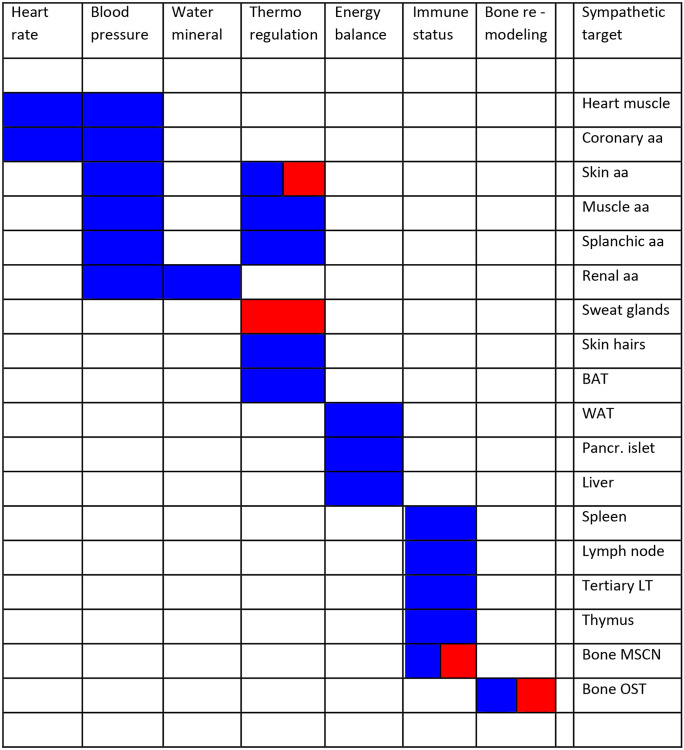
During the last 30 years, our understanding of the development and diversification of postganglionic sympathetic neurons has dramatically increased. In parallel, the list of target structures has been critically extended from the cardiovascular system and selected glandular structures to metabolically relevant tissues such as white and brown adipose tissue, lymphoid tissues, bone, and bone marrow. A critical question now emerges for the integration of the diverse sympathetic neuron classes into neural circuits specific for these different target tissues to achieve the homeostatic regulation of the physiological ends affected.
Keywords: Adiposity; Bone marrow; Cholinergic; Heartrate; Hydromineral; Hypertension; Immune status; Noradrenergic; Skeletal health; Synaptic organizer; Thermogenesis; Transdifferentiation.
© 2021. The Author(s).
Conflict of interest statement
The authors declare no competing interests.
Figures
Similar articles
-
Neurophysiological analysis of target-related sympathetic pathways--from animal to human: similarities and differences.Acta Physiol Scand. 2003 Mar;177(3):255-74. doi: 10.1046/j.1365-201X.2003.01088.x. Acta Physiol Scand. 2003. PMID: 12608996 Review.
-
The development of postganglionic sympathetic neurons: coordinating neuronal differentiation and diversification.Auton Neurosci. 2001 Dec 10;94(1-2):1-13. doi: 10.1016/S1566-0702(01)00336-8. Auton Neurosci. 2001. PMID: 11775697 Review.
-
Central neural control of thermoregulation and brown adipose tissue.Auton Neurosci. 2016 Apr;196:14-24. doi: 10.1016/j.autneu.2016.02.010. Epub 2016 Feb 23. Auton Neurosci. 2016. PMID: 26924538 Free PMC article. Review.
-
Bidirectional crosstalk between the sensory and sympathetic motor systems innervating brown and white adipose tissue in male Siberian hamsters.Am J Physiol Regul Integr Comp Physiol. 2017 Mar 1;312(3):R324-R337. doi: 10.1152/ajpregu.00456.2015. Epub 2017 Jan 11. Am J Physiol Regul Integr Comp Physiol. 2017. PMID: 28077392 Free PMC article.
-
Differential control of sympathetic outflow.Am J Physiol Regul Integr Comp Physiol. 2001 Sep;281(3):R683-98. doi: 10.1152/ajpregu.2001.281.3.R683. Am J Physiol Regul Integr Comp Physiol. 2001. PMID: 11506981 Review.
Cited by
-
Controls of Central and Peripheral Blood Pressure and Hemorrhagic/Hypovolemic Shock.J Clin Med. 2023 Jan 31;12(3):1108. doi: 10.3390/jcm12031108. J Clin Med. 2023. PMID: 36769755 Free PMC article. Review.
-
Generation of Functional and Mature Sympathetic Neurons from Human Pluripotent Stem Cells via a Neuroepithelial Route.J Mol Neurosci. 2024 Feb 15;74(1):19. doi: 10.1007/s12031-024-02196-5. J Mol Neurosci. 2024. PMID: 38358571
-
Impact of psychological stress on ovarian function: Insights, mechanisms and intervention strategies (Review).Int J Mol Med. 2025 Feb;55(2):34. doi: 10.3892/ijmm.2024.5475. Epub 2024 Dec 20. Int J Mol Med. 2025. PMID: 39704226 Free PMC article. Review.
-
Molecular and functional diversity of the autonomic nervous system.Nat Rev Neurosci. 2025 Jul 3. doi: 10.1038/s41583-025-00941-2. Online ahead of print. Nat Rev Neurosci. 2025. PMID: 40610604 Review.
References
-
- Ahrén B, Veith RC, Taborsky GJ Jr (1987) Sympathetic nerve stimulation versus pancreatic norepinephrine infusion in the dog: 1). Effects on basal release of insulin and glucagon. Endocrinology 121(1):323–331. 10.1210/endo-121-1-323 - PubMed
-
- Ali FR, Marcos D, Chernukhin I, Woods LM, Parkinson LM, Wylie LA, Papkovskaia TD, Davies JD, Carroll JS, Philpott A. Dephosphorylation of the proneural transcription factor ASCL1 re-engages a latent post-mitotic differentiation program in neuroblastoma. Mol Cancer Res. 2020;18(12):1759–1766. doi: 10.1158/1541-7786.MCR-20-0693. - DOI - PMC - PubMed
Publication types
MeSH terms
Grants and funding
LinkOut - more resources
Full Text Sources
