. 2022 Feb;44(1):293-310.
doi: 10.1007/s11357-021-00483-2. Epub 2021 Nov 10.

Mesenchymal stromal cell-derived small extracellular vesicles promote neurological recovery and brain remodeling after distal middle cerebral artery occlusion in aged rats

Affiliations

Mesenchymal stromal cell-derived small extracellular vesicles promote neurological recovery and brain remodeling after distal middle cerebral artery occlusion in aged rats

Danut-Adrian Dumbrava et al. Geroscience. 2022 Feb.

Abstract

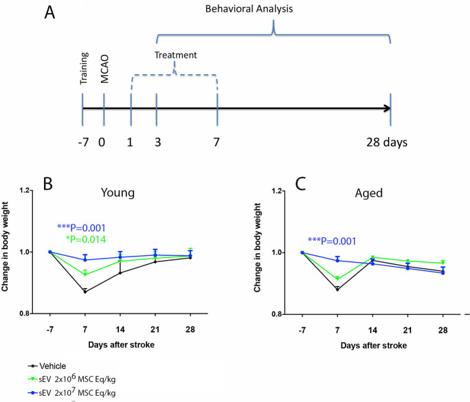
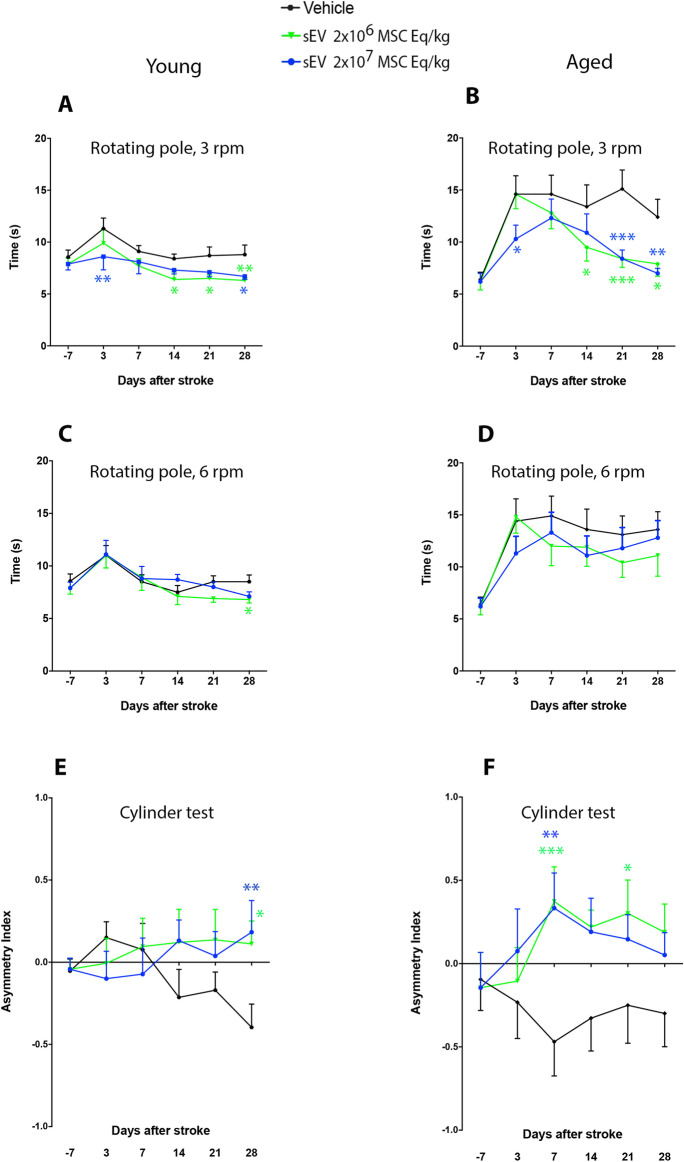
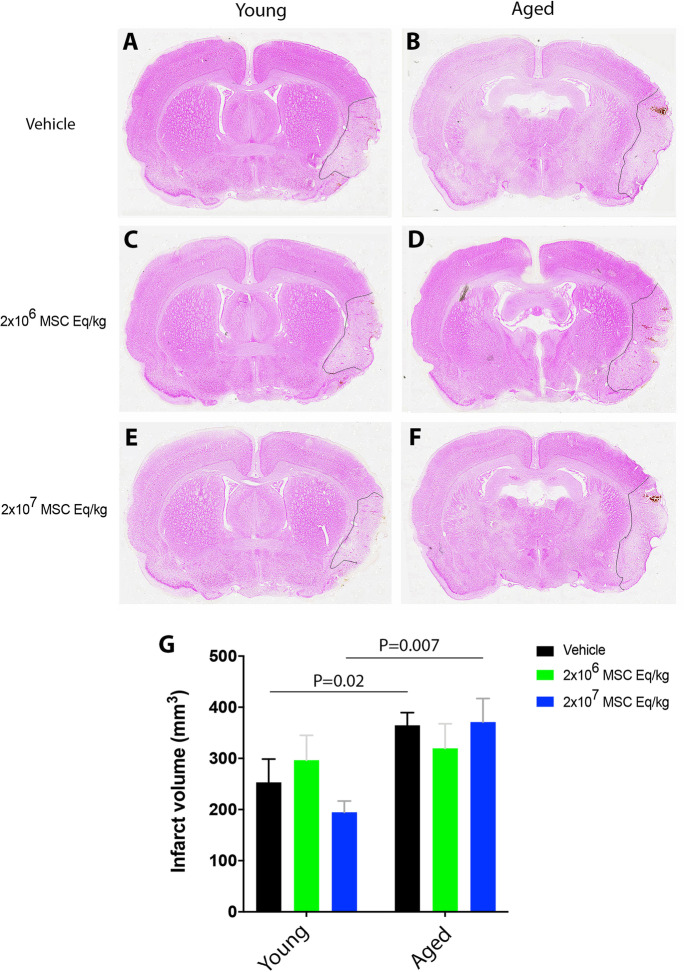
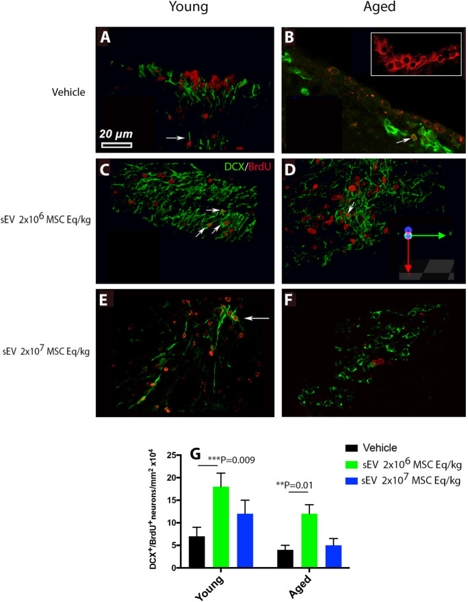
Small extracellular vesicles (sEVs) obtained from mesenchymal stromal cells (MSCs) promote neurological recovery after middle cerebral artery occlusion (MCAO) in young rodents. Ischemic stroke mainly affects aged humans. MSC-sEV effects on stroke recovery in aged rodents had not been assessed. In a head-to-head comparison, we exposed young (4-5 months) and aged (19-20 months) male Sprague-Dawley rats to permanent distal MCAO. At 24 h, 3 and 7 days post-stroke, vehicle or MSC-sEVs (2 × 10⁶ or 2 × 10⁷ MSC equivalents/kg) were intravenously administered. Neurological deficits, ischemic injury, brain inflammatory responses, post-ischemic angiogenesis, and endogenous neurogenesis were evaluated over 28 days. Post-MCAO, aged vehicle-treated rats exhibited more severe motor-coordination deficits evaluated by rotating pole and cylinder tests and larger brain infarcts than young vehicle-treated rats. Although infarct volume was not influenced by MSC-sEVs, sEVs at both doses effectively reduced motor-coordination deficits in young and aged rats. Brain macrophage infiltrates in periinfarct tissue, which were evaluated as marker of a recovery-aversive inflammatory environment, were significantly stronger in aged than young vehicle-treated rats. sEVs reduced brain macrophage infiltrates in aged, but not young rats. The tolerogenic shift in immune balance paved the way for structural brain tissue remodeling. Hence, sEVs at both doses increased periinfarct angiogenesis evaluated by CD31/BrdU immunohistochemistry in young and aged rats, and low-dose sEVs increased neurogenesis in the subventricular zone examined by DCX/BrdU immunohistochemistry. Our study provides robust evidence that MSC-sEVs promote functional neurological recovery and brain tissue remodeling in aged rats post-stroke. This study encourages further proof-of-concept studies in clinic-relevant stroke settings.

Keywords: Aging; Angiogenesis; Exosome; Ischemic stroke; Macrophage; Neurogenesis; Permanent focal cerebral ischemia; Stem/precursor cell.


Conflict of interest statement

The authors declare no competing interests.

Figures

Fig. 1
Fig. 1
Experimental design and time course of body weight changes in young and aged rats exposed to permanent distal middle cerebral artery occlusion (MCAO). A Experimental design showing the time-line of experimental interventions. Rats (n = 15 animals/group) received intravenous injections of mesenchymal stromal cell (MSC)-derived small extracellular vesicles (sEVs) at 1, 3, and 7 days post-MCAO. Motor-coordination deficits were evaluated by behavioral analysis from 3 to 28 days post-MCAO (i.e., from 2 to 27 days post-treatment onset). Rats were sacrificed after 28 days for brain tissue analysis. B, C Time course of body weight changes in young and aged rats exposed to permanent distal MCAO. Body weight decreased in young and aged vehicle-treated rats in the first 7 days post-stroke. Body weight fully recovered within 14–21 days in all groups. Note that MSC-sEVs prevented the weight loss at 7 days in young and aged rats. Data are means ± SEM values
Fig. 2
Fig. 2
MSC-derived sEVs at both doses promote post-stroke motor-coordination recovery in young and aged rats. Motor-coordination deficits in A, B the rotating pole test at 3 rpm, C, D the rotating pole test at 6 rpm, and E, F the cylinder test of A, C, E young and B, D, F aged rats exposed to permanent distal MCAO which received vehicle or sEVs (2 × 106 or 2 × 107 MSC equivalents/kg) at 1, 3, and 7 days post-stroke (n = 15 animals/group). The rotating pole test evaluates the time needed to traverse a pole rotating at a given speed (3 or 6 rpm), and the cylinder test measures the asymmetry of forelimb use, more negative data indicating reduced use of the stroke-affected left limb. Note that the test performance in young and aged rats was robustly improved by low-dose and high-dose MSC-sEVs in A, B the rotating pole test at 3 rpm and E, F the cylinder test. In comparison, C, D the rotating pole test at 6 rpm imposes an elevated task difficulty. This latter test was unable to discriminate behavioral changes between treatment groups in aged rats. Data are mean ± SEM values
Fig. 3
Fig. 3
MSC-sEVs do not influence infarct volume in young or aged rats. Infarct volume assessed by methyl green/pyronine Y staining in A, C, E young and B, D, F aged rats exposed to permanent distal MCAO, which received vehicle or sEVs (2 × 106 or 2 × 107 MSC equivalents/kg) at 1, 3, and 7 days post-stroke, followed by animal sacrifice after 28 days (n = 15 animals/group). Note that G infarct volume was significantly larger in aged than young control rats. Data are mean ± SD values. Scale bar, 1 mm
Fig. 4
Fig. 4
sEVs at both doses reduce periinfarct macrophage accumulation in aged rats. Number of ED1+ macrophages in the periinfarct cortex of A, C, E young and B, D, F aged MCAO rats assessed by immunohistochemistry. Rats received vehicle or sEVs (2 × 106 or 2 × 107 MSC equivalents/kg) at 1, 3, and 7 days post-stroke, followed by animal sacrifice after 28 days (n = 15 animals/group). Macrophages were identified using thresholds reflecting their appropriate size, which is ~ 50% larger than that of monocytes. Hence, monocytes were excluded from cell countings. Note G the exacerbated brain infiltrates of ED1+ macrophages in the brains of aged compared with young mice, which were markedly reduced by sEVs. sEVs did not influence ED1+ macrophage infiltrates in young mice. Data are mean ± SD values. Scale bar, 100 µm.
Fig. 5
Fig. 5
Low-dose sEVs reduce periinfarct microglia accumulation in young rats. Number of Iba1+ microglia in the periinfarct cortex of A, C, E young and B, D, F aged MCAO rats assessed by immunohistochemistry. Rats received vehicle or sEVs (2 × 106 or 2 × 107 MSC equivalents/kg) at 1, 3, and 7 days post-stroke, followed by animal sacrifice after 28 days (n = 15 animals/group). Note G the moderately elevated microglial recruitment in aged compared to young rats. Data are mean ± SD values. Scale bar, 50 µm
Fig. 6
Fig. 6
sEVs at both doses increase periinfarct angiogenesis in young and aged rats. Number of CD31+ brain microvessels (in green) in the periinfarct cortex of A, B, C young and D, E, F aged MCAO rats that were double labeled with the proliferation marker bromodeoxyuridine (BrdU; in red). Rats received vehicle or sEVs (2 × 106 or 2 × 107 MSC equivalents/kg) at 1, 3, and 7 days post-stroke (n = 15 animals/group). For the labeling of proliferating cells, BrdU (50 mg/kg body weight) was intraperitoneally administered daily from 8 to 18 days, followed by animal sacrifice after 28 days. Note G the dose-dependent stimulation of angiogenesis by sEVs in young and aged rats. Arrows depicting CD31+/BrdU+ double labeled cells. Data are mean ± SD values. Scale bar, 20 µm.
Fig. 7
Fig. 7
Low-dose sEVs stimulates post-stroke neurogenesis adjacent to the subventricular zone of young and aged rats. Number of newborn neurons adjacent to the subventricular zone (SVZ) of A, C, E young and B, D, F aged MCAO rats expressing the immature neuronal marker doublecortin (DCX; in green) that were double labeled by the proliferation marker BrdU (in red). Rats received vehicle or sEVs (2 × 106 or 2 × 107 MSC equivalents/kg) at 1, 3, and 7 days post-stroke (n = 15 animals/group). For cell proliferation analysis, BrdU (50 mg/kg body weight) was intraperitoneally administered daily from 8 to 18 days, followed by animal sacrifice after 28 days. Note G that low-dose sEVs increased endogenous neurogenesis in young and aged rats, whereas high-dose sEVs did not have any significant effect. Note that most DCX+ cells did not colocalize with BrdU+ nuclei in the SVZ of vehicle-treated rats (A, B; arrows). Instead, the BrdU+ nuclei were distributed mainly in a “pinwheel” configuration adjacent to the periventricular epithelium (B, inset). Arrows in (CE depicting DCX+/BrdU+ cells. Data are mean ± SD values. Scale bar, 20 µm

References

    1. Xin H, Li Y, Buller B, Katakowski M, Zhang Y, Wang X, et al. Exosome-mediated transfer of miR-133b from multipotent mesenchymal stromal cells to neural cells contributes to neurite outgrowth. Stem Cells. 2012;30(7):1556–1564. doi: 10.1002/stem.1129. - DOI - PMC - PubMed
    1. Xin H, Li Y, Cui Y, Yang JJ, Zhang ZG, Chopp M. Systemic administration of exosomes released from mesenchymal stromal cells promote functional recovery and neurovascular plasticity after stroke in rats. J Cereb Blood Flow Metab. 2013;33(11):1711–1715. doi: 10.1038/jcbfm.2013.152. - DOI - PMC - PubMed
    1. Doeppner TR, Bähr M, Hermann DM, Giebel B. Concise review: extracellular Vesicles overcoming limitations of cell therapies in ischemic stroke. Stem Cells Transl Med. 2017;6(11):2044–2052. doi: 10.1002/sctm.17-0081. - DOI - PMC - PubMed
    1. Doeppner TR, Herz J, Görgens A, Schlechter J, Ludwig AK, Radtke S et al. Extracellular vesicles improve post-stroke neuroregeneration and prevent postischemic immunosuppression. Stem Cells Transl Med. 2015;4(10):1131–1143. 12x10 MSC equivalents/kg966/sctm.2015–0078. - PMC - PubMed
    1. Yáñez-Mó M, Siljander PR, Andreu Z, Zavec AB, Borràs FE, Buzas EI, et al. Biological properties of extracellular vesicles and their physiological functions. J Extracell Vesicles. 2015;4:27066. doi: 10.3402/jev.v4.27066. - DOI - PMC - PubMed

Publication types

LinkOut - more resources