. 2021 Dec;78(6):1818-1828.
doi: 10.1161/HYPERTENSIONAHA.121.17926. Epub 2021 Nov 1.

Noninvasive Biomarkers for Cardiovascular Dysfunction Programmed in Male Offspring of Adverse Pregnancy

Affiliations

Noninvasive Biomarkers for Cardiovascular Dysfunction Programmed in Male Offspring of Adverse Pregnancy

Rama Lakshman et al. Hypertension. 2021 Dec.

Abstract

[Figure: see text].

Keywords: biomarkers; cardiovascular diseases; fetal hypoxia; oxidative stress; pregnancy.


Figures

Figure 1.
Figure 1.
Origins of arterial blood pressure (BP) and heart rate (HR) variability. Acute changes in BP are restored via the baroreflex. A change in BP is detected by arterial baroreceptors, which signal to the medulla. This triggers a compensatory change in HR, and thus cardiac output [CO], via reciprocal modulation of sympathetic and vagal activity to the cardiac sinoatrial node. There is also a change in sympathetic outflow to peripheral arterioles, resulting in a compensatory change in total peripheral vascular resistance (TPR). Very-low-frequency (VLF; red box) blood pressure variability (BPV) occurs due to myogenic responses creating a VLF oscillation in peripheral arteriolar tone and thus TPR. The VLF BPV activates the baroreflex leading to compensatory VLF HRV. Low-frequency (LF) BPV and HRV originate from baroreflex loop resonance (red box). At the resonant frequency, the time delay in this negative feedback loop means the input and output are in phase, generating self-sustained oscillations. High-frequency (HF) BPV and HRV correspond to respiration (red box). The mechanical changes during respiration lead to HF BP oscillations (inspiration lowers intrathoracic pressure, leading to increased venous return, stroke volume [SV], and, therefore, CO), which then activate the baroreflex to produce compensatory HR oscillations.
Figure 2.
Figure 2.
Experimental design. Days gestational age (dGA) for the induction of prenatal hypoxia, and MitoQ intervention, weaning at 21 postnatal days (dPN), and cardiovascular assessment at 4.5 postnatal months (mPN).
Figure 3.
Figure 3.
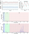
Cardiovascular recording analysis. Representative LabChart recording of continuous, pulsatile arterial blood pressure (red trace) and femoral blood flow (blue trace) from a normoxic rat (A). Expanded section of the blood pressure trace (B); the peak height (systolic blood pressure) and interpeak interval are noted. Values for systolic blood pressure and interpeak interval were plotted against time and fast Fourier transformed into the frequency domain to produce blood pressure variability (BPV; C) and heart rate variability (HRV; D) power spectra, respectively. HF indicates high frequency; LF, low frequency; and VLF, very low frequency.
Figure 4.
Figure 4.
Baseline cardiovascular function. Mean arterial pressure (A) and mean heart rate (B) in male offspring from normoxic (N; white, n=8 and 10, respectively), hypoxic (H; gray, n=6 and 9, respectively), hypoxic+MitoQ (HM; red, n=6 and 8, respectively), and normoxic+MitoQ (NM; blue, n=4 and 6, respectively) pregnancies. Data are mean±SEM. Two-way ANOVA for the effect of hypoxia (*P<0.05) and the effect of MitoQ (†P<0.05). BPM indicates beats per minute.
Figure 5.
Figure 5.
Blood pressure variability (BPV). Very-low-frequency (VLF; A) and low-frequency (LF; B) blood pressure variability (BPV) in male offspring from normoxic (N; white, n=8), hypoxic (H; gray, n=6), hypoxic+MitoQ (HM; red, n=6), and normoxic+MitoQ (NM; blue, n=4) pregnancies. Data are mean±SEM. Two-way ANOVA for the effect of hypoxia (*P<0.05) and the effect of MitoQ (†P<0.05).
Figure 6.
Figure 6.
Heart rate variability. The SD of the interbeat intervals (SDNNs; A), very-low-frequency (VLF) heart rate variability (HRV; B), low-frequency (LF)/high-frequency (HF) ratio (C) in male offspring from normoxic (N; white, n=10), hypoxic (H; gray, n=9), hypoxic+MitoQ (HM; red, n=8), and normoxic+MitoQ (NM; blue, n=6) pregnancies, and normalized LF (black bars) and HF (white bars) HRV (D). Data are mean±SEM. Two-way ANOVA for the effect of hypoxia (*P<0.05) and the effect of MitoQ (†P<0.05).

References

    1. Spiroski AM, Niu Y, Nicholas LM, Austin-Williams S, Camm EJ, Sutherland MR, Ashmore TJ, Skeffington KL, Logan A, Ozanne SE, et al. . Mitochondria antioxidant protection against cardiovascular dysfunction programmed by early-onset gestational hypoxia. FASEB J. 2021;35:e21446. doi: 10.1096/fj.202002705R - PMC - PubMed
    1. Demicheva E, Crispi F. Long-term follow-up of intrauterine growth restriction: cardiovascular disorders. Fetal Diagn Ther. 2014;36:143–153. doi: 10.1159/000353633 - PubMed
    1. Giussani DA, Davidge ST. Developmental programming of cardiovascular disease by prenatal hypoxia. J Dev Orig Health Dis. 2013;4:328–337. doi: 10.1017/S204017441300010X - PubMed
    1. Gluckman PD, Hanson MA, Cooper C, Thornburg KL. Effect of in utero and early-life conditions on adult health and disease. N Engl J Med. 2008;359:61–73. doi: 10.1056/NEJMra0708473 - PMC - PubMed
    1. Niu Y, Kane AD, Lusby CM, Allison BJ, Chua YY, Kaandorp JJ, Nevin-Dolan R, Ashmore TJ, Blackmore HL, Derks JB, et al. . Maternal Allopurinol prevents cardiac dysfunction in adult male offspring programmed by chronic hypoxia during pregnancy. Hypertension. 2018;72:971–978. doi: 10.1161/HYPERTENSIONAHA.118.11363 - PMC - PubMed

Publication types