. 2021 Nov 12;7(46):eabj9846.
doi: 10.1126/sciadv.abj9846. Epub 2021 Nov 10.

Phase transition specified by a binary code patterns the vertebrate eye cup

Affiliations

Phase transition specified by a binary code patterns the vertebrate eye cup

Revathi Balasubramanian et al. Sci Adv. .

Abstract

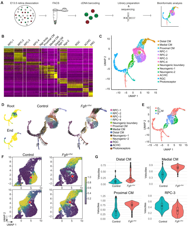
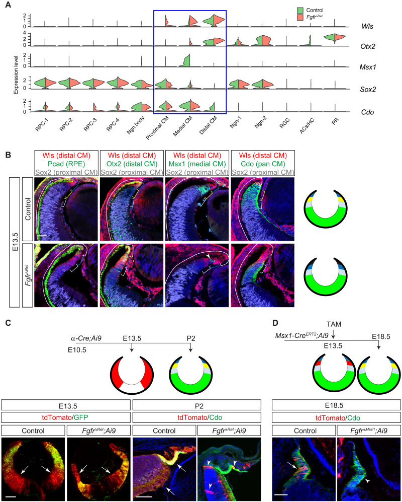
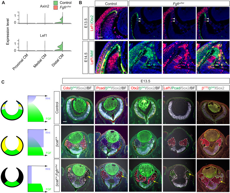
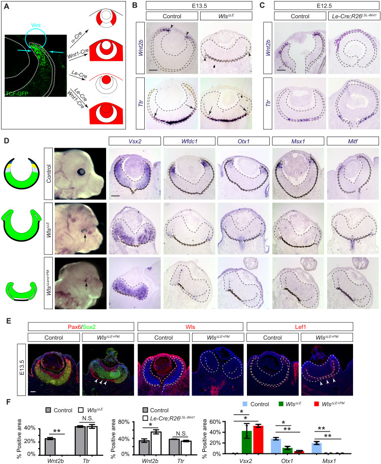
The developing vertebrate eye cup is partitioned into the neural retina (NR), the retinal pigmented epithelium (RPE), and the ciliary margin (CM). By single-cell analysis, we showed that fibroblast growth factor (FGF) signaling regulates the CM in its stem cell–like property of self-renewal, differentiation, and survival, which is balanced by an evolutionarily conserved Wnt signaling gradient. FGF promotes Wnt signaling in the CM by stabilizing β-catenin in a GSK3β-independent manner. While Wnt signaling converts the NR to either the CM or the RPE depending on FGF signaling, FGF transforms the RPE to the NR or CM dependent on Wnt activity. The default fate of the eye cup is the NR, but synergistic FGF and Wnt signaling promotes CM formation both in vivo and in human retinal organoid. Our study reveals that the vertebrate eye develops through phase transition determined by a combinatorial code of FGF and Wnt signaling.


Figures

Fig. 1.
Fig. 1.. Genetic ablation of FGFRs disrupted CM development.
(A) Evolutionary conservation of peripheral ocular structures. The vertebrate eye cup is partitioned into the NR, the RPE, and the CM, the latter of which gives rise to the CB and the iris. This resembles the Drosophila eye where the NR is shielded by the pigment rim (PR), the head capsule (HC), and the subretinal pigment (SRP). (B) Deletion of FGFRs in the peripheral retina abolished FGF signaling as indicated by the loss of pERK, Etv5, and Spry2 expression. (C) NR domain (dotted lines) marked by Vsx2, Sox2, Pax6, NICD, Gli1, Sfrp2, and Atoh7 expression was reduced in FgfrΔRet mutants. (D) FgfrΔRet mutant retina displayed ectopic expression of Mitf, Pcad, and Cx43 (dotted lines) but down-regulated Wfdc1, indicating loss of the CM domain. (E) FgfrΔRet mutant animals exhibited dysmorphic CB (arrows) and iris hypoplasia (arrowheads) at P7 and aniridia (white arrowheads) in adulthood. Scale bars, 100 μm.
Fig. 2.
Fig. 2.. Single-cell analysis showed that FGF signaling regulates self-renewal and differentiation of CM progenitors.
(A) Schematic diagram of scRNAseq. (B) Heatmap of differential gene expression in single-cell clusters. RPC, retinal progenitor cell; RGC, retinal ganglion cell; AC, amacrine cell; HC, horizontal cell; PR, photoreceptor. (C) UMAP representation of single-cell clusters. (D) Left: Diffusion analysis of RNA velocities identified the root and the end of cell differentiation as represented by high-density regions (dark green) after forward and reverse Markov processes. Right: Cell differentiation trajectories were revealed by the velocity field projected on the UMAP plot. Arrows indicate local RNA velocities on a regular grid. (E) State of the cell cycle represented on the UMAP plot. (F) Single-step transition probabilities from the starting cells (red square) to neighboring cells showed the bias of CM progenitors in FgfrΔRet mutants toward differentiation against self-renewal. (G) Quantification of single-cell velocities in each cluster showed the accelerated differentiation and reduced proliferation of CM progenitor cells.
Fig. 3.
Fig. 3.. FGF signaling is required for CM subdivision and survival.
(A) Violin plots showed that the CM can be subdivided by the overlapping expression of Wls, Otx2, Msx1, Sox2, and Cdo, all of which were dysregulated in FgfrΔRet mutants. (B) Immunostaining confirmed that the in silico clustering of CM cells matches the spatial separation of CM subdomains. Aberrant invasion of Pcad, expansion of Wls and Otx2, loss of Msx1, and reduction in Sox2 and Cdo in FgfrΔRet mutants demonstrated CM differentiation defects. Brackets indicate the domain of the RPE and the distal, medial, and proximal CM, corresponding to black, dark blue, yellow, and light blue regions, respectively, in diagrams on the right. The NR is indicated in green. Arrowhead marks residual wild-type cells still expressing Msx1. Scale bar, 50 μm. (C) At E13.5, although Pax6 α-Cre was expressed only in the peripheral retina indicated by its GFP reporter, it has already activated tdTomato expression (arrows) from the Ai9 Cre reporter throughout the retina. At P2, these tdTomato+ progenies remained in control retinae, but only a few (arrowheads) were left in FgfrΔRet mutants. The tdTomato expression is indicated in red in diagrams. Scale bars, 100 μm. (D) CM cells were pulse-labeled by tamoxifen induction of Msx1-CreERT2 at E13.5 and detected at E18.5 by tdTomato expression from the Ai9 Cre reporter. Although these cells remained in the control CM identified by Cdo expression (arrow), they had largely disappeared in the FgfrΔRet mutant (arrowhead), suggesting cell survival defects. Scale bar, 50 μm.
Fig. 4.
Fig. 4.. CM development is dependent on the dosage of FGF signaling.
(A) scRNAseq analysis revealed a nested pattern of Fgf3, Fgf9, and Fgf15 expression in the retina. (B) By reducing the dosage of Fgf ligands, Fgf9ΔRet and Fgf3/9ΔRet exhibited progressive expansion of the distal CM/RPE marker Mitf, reduction in the proximal CM/NR marker Vsx2, and loss of pan-CM marker Wfdc1 and medial CM marker Msx1. (C) Overexpression of Fgf8 in Fgf8OE mutants stimulated pERK and FGF-responsive gene Spry2 in the presumptive RPE territory, which was transformed to NR as indicated by Atoh7 expression. Reducing the strength of FGF signaling by deleting Fgfr1 and Fgfr2 in FgfrΔRet;Fgf8OE mutants down-regulated pERK. This prevented expression of Atoh7 but induced the CM markers Otx1 and Msx1, indicating the transformation to the CM fate. The Cre expression in the RPE territory was indicated by GFP expression from the α-Cre driver. Scale bars, 100 μm. (D) Relative area expressing each marker gene was normalized against the entire inner (left) or outer (right) eye cup, and the statistical significance was evaluated using one-way analysis of variance (ANOVA) test. *P < 0.01, **P < 0.001, and ***P < 0.0001; n = 3 for all markers.
Fig. 5.
Fig. 5.. FGF signaling determines the CM fate by promoting Wnt signaling.
(A) Violin plots showed down-regulation of Wnt response genes Lef1 and Axin2 in the FgfrΔRet mutant transcriptome. (B) In mosaic analysis, FgfrΔRet mutant cells acquired ectopic expression of Otx2 and Mitf at the expense of Lef1. In contrast, the remaining wild-type cells identified by the lack of Otx2 and Mitf expression still maintained Lef1 expression. Scale bar, 50 μm. (C) Constitutive activation of Wnt signaling by deleting the β-cateninSer45Ser45) motif transformed βcatCA retinae to the CM as indicated by Cdo expression. Further deletion of FGFRs in FgfrΔRetcatCA mutant retinae converts them to the RPE as indicated by ectopic expression of Pcad and Otx2 as well as the appearance of pigmentation (arrows). Both the level of Lef1 and the amount of β-catenin detected by the β-catenin C-terminal antibody (βCTD) arose in βcatCA retinae but declined in FgfrΔRetcatCA mutants, showing that the Wnt–β-catenin signaling is dependent on FGF signaling. The mutant regions are marked by dotted lines. Scale bar, 100 μm.
Fig. 6.
Fig. 6.. Paracrine Wnt signaling patterns the CM and the distal RPE.
(A) Potential source of the Wnt signaling gradient revealed by the TCF-GFP reporter may be targeted by α-Cre for retina, Wnt1-Cre for periocular mesenchyme, and Le-Cre for the lens ectoderm (the surface epithelium and the lens). (B) Ablation of Wnt transporter Wls in the lens ectoderm specifically disrupted the proximal RPE differentiation in WlsΔLE mutants as indicated by loss of Wnt2b but not the proximal RPE marker Ttr. (C) Overexpression of Wnt1 in the lens ectoderm also affected the proximal RPE by expanding the domain of Wnt2b expression in Le-Cre;R26LSL-Wnt1 eye cup without affecting Ttr expression. (D) CM was lost in WlsΔLE mutants as shown by the expansion of Vsx2 and the absence of Wfdc1, Otx1, Msx1, and Mitf. The band of the RPE was further diminished in WlsΔLE+ΔPM mutants. (E) WlsΔLE+ΔPM mutant eyes lost Wls and Lef1 in both the lens ectoderm and the periocular mesenchyme, leaving only a small band of Lef1 expression next to the Pax6-expressing RPE (arrowheads). Scale bars, 100 μm. (F) Relative area expressing Wnt2b, Ttr, or Vsx2 was normalized against the entire RPE region, while the Otx1- or Msx1-expressing area was normalized against the NR region. Student’s t test for Wnt2b and Ttr and one-way ANOVA test for Vsx2, Otx1, and Msx1. *P < 0.05 and **P < 0.01; n = 2 for all markers. N.S., not significant.
Fig. 7.
Fig. 7.. FGF and Wnt induce phase transition to generate the CM.
(A) Deletion of either β-catenin alone (βcatΔRet) or β-catenin and Fgfrs together (FgfrΔRetcatΔRet) transformed the distal retina into the NR as indicated by loss of Cdo and Msx1, lack of Otx2 and Pcad, and expansion of Sox2 (all at 100% in the β-catenin or β-catenin/pERK–deficient regions marked by dotted lines; n = 3). (B) Although overexpression of Wnt1 in Wnt1OE embryos did not produce any phenotype in the retina, when combined with overexpression of Fgf8, it transformed the RPE domain of the Wnt1OE;Fgf8OE eye cup into the CM as indicated by the up-regulation of the CM-specific genes Otx1 and Cdo and the lack of the RPE marker Mitf and the NR marker Atoh7. (C) Untreated hiPSC organoid culture contained few MSX1+ or CDO+ CM-like cells. Addition of the 3 μM WNT agonist CHIR99021 significantly expanded the number of MSX1+ and CDO+ cells. (D) Quantification of the MSX1+ and CDO+ area as the percentage of the total organoid culture. (E) FGF and Wnt signaling promotes the abrupt and reversible transition of eye cup progenitors into three phases: the NR, the CM, and the RPE. Our study showed that the NR can be transformed by constitutive activation of Wnt signaling (βcatCA) into the CM, which is converted to the RPE after the loss of FGF signaling (FgfrΔ). The RPE reverts back to the CM by titrating FGF signaling (Fgf8OE;FgfrΔ). Otherwise, ablation of Wnt signaling (βcatΔ) or overexpression of Fgf8 (Fgf8OE) can turn the RPE into the NR, which may transition further into the CM after overexpression of Wnt1 (Wnt1OE). Scale bars, 100 μm.

References

    1. Chow R. L., Lang R. A., Early eye development in vertebrates. Annu. Rev. Cell Dev. Biol. 17, 255–296 (2001). - PubMed
    1. Fuhrmann S., Zou C., Levine E. M., Retinal pigment epithelium development, plasticity, and tissue homeostasis. Exp. Eye Res. 123, 141–150 (2014). - PMC - PubMed
    1. Tomlinson A., Patterning the peripheral retina of the fly: Decoding a gradient. Dev. Cell 5, 799–809 (2003). - PubMed
    1. Fujimura N., Taketo M. M., Mori M., Korinek V., Kozmik Z., Spatial and temporal regulation of Wnt/β-catenin signaling is essential for development of the retinal pigment epithelium. Dev. Biol. 334, 31–45 (2009). - PubMed
    1. Westenskow P., Piccolo S., Fuhrmann S., β-Catenin controls differentiation of the retinal pigment epithelium in the mouse optic cup by regulating Mitf and Otx2 expression. Development 136, 2505–2510 (2009). - PMC - PubMed