. 2021 Nov 10;13(619):eabh1314.
doi: 10.1126/scitranslmed.abh1314. Epub 2021 Nov 10.

Selective targeting of NaV1.7 via inhibition of the CRMP2-Ubc9 interaction reduces pain in rodents

Affiliations

Selective targeting of NaV1.7 via inhibition of the CRMP2-Ubc9 interaction reduces pain in rodents

Song Cai et al. Sci Transl Med. .

Abstract

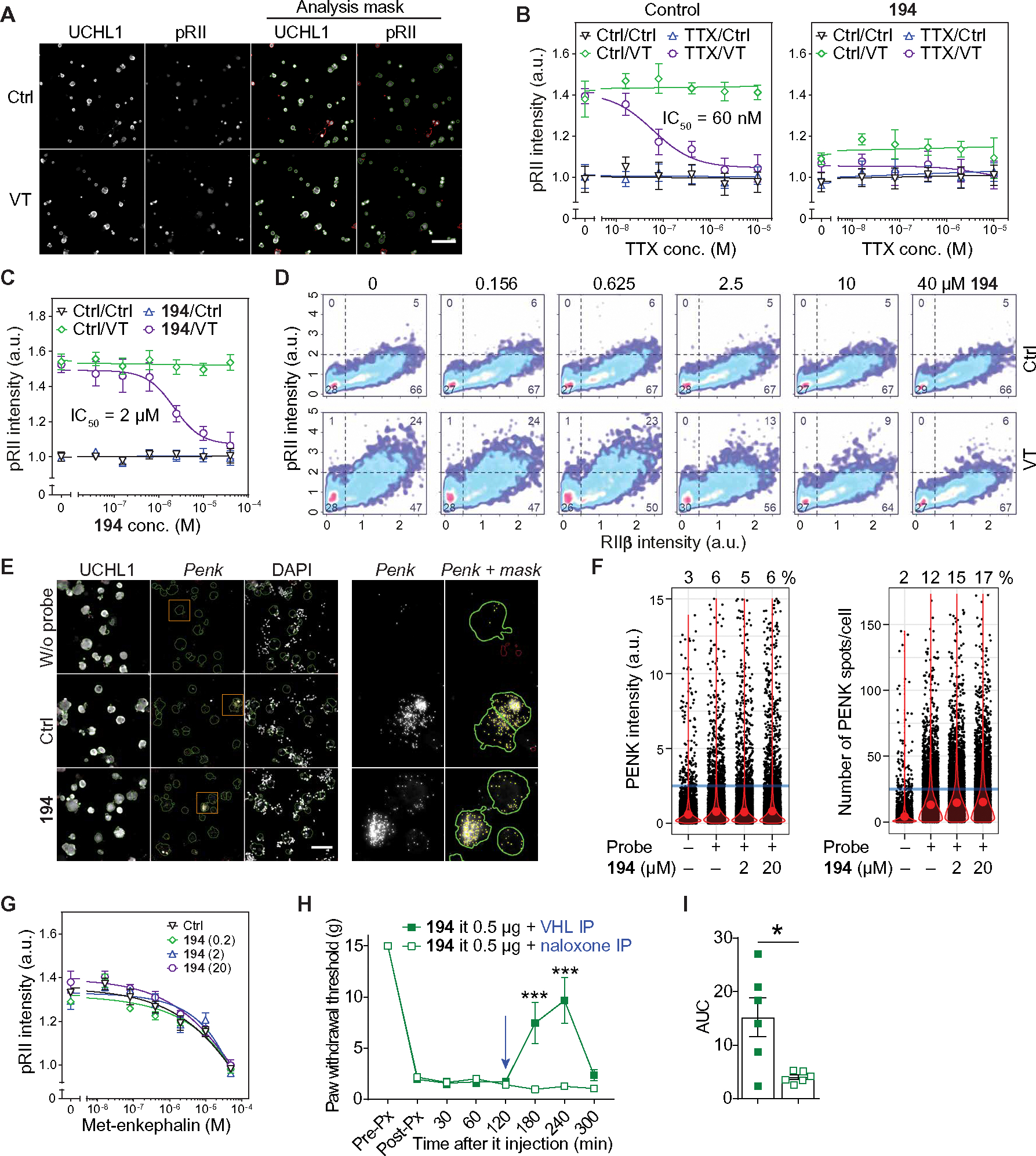
The voltage-gated sodium NaV1.7 channel, critical for sensing pain, has been actively targeted by drug developers; however, there are currently no effective and safe therapies targeting NaV1.7. Here, we tested whether a different approach, indirect NaV1.7 regulation, could have antinociceptive effects in preclinical models. We found that preventing addition of small ubiquitin-like modifier (SUMO) on the NaV1.7-interacting cytosolic collapsin response mediator protein 2 (CRMP2) blocked NaV1.7 functions and had antinociceptive effects in rodents. In silico targeting of the SUMOylation site in CRMP2 (Lys374) identified >200 hits, of which compound 194 exhibited selective in vitro and ex vivo NaV1.7 engagement. Orally administered 194 was not only antinociceptive in preclinical models of acute and chronic pain but also demonstrated synergy alongside other analgesics—without eliciting addiction, rewarding properties, or neurotoxicity. Analgesia conferred by 194 was opioid receptor dependent. Our results demonstrate that 194 is a first-in-class protein-protein inhibitor that capitalizes on CRMP2-NaV1.7 regulation to deliver safe analgesia in rodents.


Conflict of interest statement

Competing interests: R.K., M.K., and V.G. are cofounders of Regulonix LLC, a company developing nonopioid drugs for chronic pain. In addition, R.K., M.K., R.C., and V.G. have patents US10287334 (non-narcotic CRMP2 peptides targeting sodium channels for chronic pain) and US10441586 (SUMOylation inhibitors and uses thereof) issued to Regulonix LLC. The other authors declare that they have no competing interests. R.K. and A.M. are cofounders of ElutheriaTx Inc., a company developing gene therapy approaches for chronic pain.

Figures

Fig. 1.
Fig. 1.. Identification and characterization of compound 194 as an inhibitor of CRMP2-Ubc9 interaction and inhibitor of NaV1.7 currents in sensory neurons from multiple species.
(A) Ribbon representation of the tripartite interaction between CRMP2 (dark pink), Ubc9 (orange), and SUMO2 (black). Key residues involved in the interaction are outlined as follows: V371 of CRMP2 interacts with A131 of Ubc9, and R440 of CRMP2 forms a salt bridge with E132 of Ubc9, and the K374 residue of CRMP2 to which the SUMO protein is positioned for covalent attachment. (B) Close-up representation of the best docking pose of 194 (green) near the interface between CRMP2 and Ubc9. (C) Structure of 002 and 145 and of the lead compound 194, which presents with features from both core pharmacophores. (D) One-dimensional 1H saturation transfer difference nuclear magnetic resonance (STD NMR) showing resonance difference spectrum for CRMP5, CRMP2, and Ubc9 in the presence of 194. Regions that yielded an STD signal in the presence of 194 are indicated with #. ppm, parts per million. (E) Microscale thermophoresis of nitrilotriacetic acid (NTA)–labeled CRMP2 interaction with Ubc9 in the absence or presence of increasing concentrations of 194. Data were fitted with a one-site binding model. (F) Representative sodium current traces recorded from small-sized DRG neurons, incubated overnight with 194 (5 μM) with or without the NaV1.7-selective inhibitor ProTx-II (24) (5 nM, 5 to 15 min), in response to depolarization steps from −70 to +60 mV from a holding potential of −60 mV. Summary of current-voltage curves (G) and normalized peak (H) currents (picoamperes/picoFarads) from small (<20 μm) (i) or large (~38 to 51 μm). p = 0.0020, 0.0005, and 0.0300 for DMSO versus ProTx-II, 194, and 194 + ProTx-II, respectively (one Way-ANOVA; see table S1). (I) DRG neurons as indicated (n = 10 to 12 cells per group for small and n = 9 to 11 cells per group for large). (J) Concentration curve illustrating inhibition of NaV1.7 currents by 194 with an inhibitory constant IC50 of 1.2 μM (n = 12 to 25 cells per concentration). (K) Percentage inhibition of NaV1.x-mediated currents in HEK293 cell lines expressing NaV1.x subunits (yellow boxes) or rat DRGs (NaV1.7–9; gray boxes) after overnight treatment with 20 μM 194. NaV1.7 was isolated with TTX (17), NaV1.8 was isolated with 500 nM selective blocker A803467 (70), whereas NaV1.9 was isolated with post hoc subtraction techniques described previously (17, 18). (L) Bar graph of normalized peak NaV1.7 current densities of mouse, pig, and human DRG neurons treated overnight with 5 μM 194 (n = 9 to 12 cells per condition). (M) Bar graph of normalized peak NaV1.7 current densities of DRGs from wild-type and CRMP2K374A/K374A mice neurons treated overnight with 5 μM 194 (n > 15 cells per condition). Complete sample size and statistical information are provided in table S1. Error bars indicate mean ± SEM.
Fig. 2.
Fig. 2.. 194 blocks CRMP2 SUMOylation promoting recruitment of endocytosis-related proteins and internalization of NaV1.7.
(A) Representative immunoblot and (B) quantification of SUMO1 immunoprecipitates from Cath.-a-differentiated (CAD) cells treated overnight with 194 (5 μM) or transfected with the SUMO-null CRMP2-K374A plasmid and probed with anti-CRMP2 or anti-cdk5 antibodies (n = 3 independent samples). Ab, antibody. * denotes p = 0.0095 and 0.0047 comparing wildtype to 194 or K374A, respectively (Kruskal-Wallis test). (C) Representative immunoblot and (D) quantification of CRMP2 immunoprecipitates from CAD cells treated overnight with 194 (5 μM) or transfected with the SUMO-null CRMP2-K374A plasmid and probed with antibodies against NaV1.7, CaV2.2, βIII-tubulin, and endocytic proteins Nedd4–2, Eps15, Numb, and CRMP2 as control (n = 3 independent samples). * denotes p = 0.0014 and 0.0242 comparing NaV1.7 binding to CRMP2 between wildtype and 194 or K374A respectively; p = 0.0507 and 0.0188 comparing Nedd4–2 binding to CRMP2 between wildtype and 194 or K374A respectively; and p = 0. 0.0175 and 0.0507 comparing Eps15 binding to CRMP2 between wildtype and 194 or K374A respectively (Kruskal-Wallis test). (E) Representative immunoblot and (F) quantification of the biotinylated-fraction of NaV1.7 in CAD cells treated overnight with 194 (5 μM) or transfected with CRMP2-K374A construct (n = 3 independent samples). * denotes p = 0.0100 and 0.0062 comparing NaV1.7 surface expression between wildtype and 194 or K374A, respectively (Kruskal-Wallis test). (G) Representative images of rat DRG cultures labeled with an extracellular epitope antibody against NaV1.7. (H) Quantification of normalized surface expression of NaV1.7 per neuron (n = 37 to 45 cells). * denotes p <0.0001 between wildtype and 194 (Mann Whitney test). (I) Representative traces and (J) graph of normalized peak Na+ current density (pA/pF) from rat DRG neurons treated with 194 (5 μM) overnight with or without pretreatment (30 min before 194 application) with 30 μM CME inhibitor PitStop (n = 16 to 19 cells per condition). * denotes p = 0.0108 comparing DMSO and 194 (One-way ANOVA with Tukey’s post doc test). Complete sample size and statistical information are provided in table S1. Error bars indicate mean ± SEM.
Fig. 3.
Fig. 3.. 194 inhibits CRMP2 SUMOylation, decreases the presynaptic fraction of NaV1.7 in vivo, and reduces sEPSC and action potentials in the lumbar horn of the spinal cord.
(A) Representative immunoblot and (B) quantification of SUMO1 immunoprecipitates from spinal cord lysates of rats prepared 2 hours after intrathecal administration with 194 (0.5 μg in 5 μl) plasmid and probed with anti-CRMP2 antibody (n = 6 independent samples). βIII-Tubulin was used as a loading control. * denotes p = 0.0022 comparing SUMO IP between DMSO and 194 (Mann Whitney test). (C) Representative immunoblots of presynaptic fractions of lumbar dorsal horn samples from rats after intrathecal administration with 194 (0.5 μg in 5 μl) probed with antibodies against NaV1.7, the presynaptic marker synaptophysin (Syn.), and membrane-associated protein flotillin (loading control). PSD, postsynaptic density. (D) Quantification of NaV1.7 in the presynaptic fraction (n = 4 rats per condition). * denotes p = 0.0002 comparing relative NaV1.7 presynaptic localization between DMSO and 194 (Mann Whitney test). (E) Representative spontaneous excitatory postsynaptic potential (sEPSC) recordings of lumbar dorsal horn neurons perfused with 0.1% dimethyl sulfoxide (DMSO) or 194 (5 μM). Summary of (F) amplitudes and (G) frequencies of sEPSCs for both groups (n = 11 cells per condition). * denotes p = 0.0018 comparing mean frequency between wildtype and 194 (Mann Whitney test). (H) Representative action potential recordings of lumbar dorsal horn neurons perfused with 0.1% DMSO or 194 (5 μM). (I) Summary of the number of action potentials recorded as a function of current injected (n = 6 to 7 cells per condition). * denotes p <0.01 comparing # of action potentials, at current injections of >15 pA, between DMSO and 194 (Mann Whitney test). Complete sample size and statistical information are provided in table S1. Error bars indicate mean ± SEM.
Fig. 4.
Fig. 4.. 194 is antinociceptive when administered intrathecally or orally and produces synergistic antiallodynic effects with other analgesics.
Time course (A) and quantification (B) of paw withdrawal threshold (PWT) of male rats that underwent spared nerve injury (SNI). At 14 days after SNI, 194 was administered either intrathecally (0.5 μg/5 μl) or in the paw (2.0 μg/50 μl, intraplantar). Time course (C) and quantification (D) of paw withdrawal threshold (PWT) of male rats that underwent spinal nerve ligation (SNL) surgery. At 21 days after SNL injury, decreased PWTs were noted. Rats were orally (po) administered 2 or 10 mg/kg of 194 (n = 6 rats per group). Sustained administration of 194 [1 mg/kg per day, subcutaneously (sc)] was achieved via osmotic minipump, for 14 days, and tactile (E and F) and thermal (G and H) hypersensitivity induced by SNL was assessed. Behavior was tested at multiple time points indicated in the bar graph. Time course (I) and quantification (J) of PWTs of male rats that underwent SNL surgery and administered doses of morphine (MOR; 2 or 6 mg/kg, po) alone or in combination with vehicle or 194 (0.5 mg/kg, po) (n = 6 rats per group). Time course (K) and quantification (L) of PWTs of male rats that underwent SNL surgery and administered doses of gabapentin (GBP; 3 or 10 mg/kg, po) alone or in combination with vehicle or 194 (0.5 mg/kg, po) (n = 6 rats per group). Complete sample size and statistical information are provided in table S1. Error bars indicate mean ± SEM.
Fig. 5.
Fig. 5.. Antinociceptive mechanism of action of 194 involves PENK up-regulation and PKA-II inhibition downstream of opioid receptor engagement.
(A) Representative HCS microscopy images of veratridine (VT) (100 μM, 5 min) or control (Ctrl, 0.2% DMSO) stimulated rat DRG neurons labeled with UCHL1 to identify neurons and pRII to quantify PKA-II signaling. Green/red encircled neurons indicate automatically selected/rejected objects, respectively. Scale bar, 200 μm. (B) DRG neurons were cultured in the absence (0.2 % DMSO) or presence of 194 (20 μM in 0.2% DMSO). After 12 hours, cells were pretreated with increasing doses of TTX (0 to 10 μM, 5 min) and then tested for veratridine-induced (10 μM, 5 min) PKA-II activity as measured by anti–phospho-RII staining as a proxy for PKA-II activity. a.u., artbitrary units. (C) DRG neurons were pretreated for 6 hours with increasing doses of 194 (0 to 40 μM), followed by stimulation with VT (100 μM, 3 min), and again PKA-II activity as measured by anti–phospho-RII staining as a proxy for PKA-II activity. Values represent means ± SEM; n = 4 replicate experiments; >2500 neurons per condition. (D) Single cell–based density plots of pRII/RIIβ-labeled DRG neurons shown in (C). Dashed lines indicate the gating threshold to discriminate activated (those with increased pRII intensity) and nociceptive [those with increased RIIβ(+)] intensity, neurons with the numbers indicating the relative percentage of cells in the respective quadrant. Combined data of n = 4 experiments with a total of >2500 neurons per condition. (E) HCS microscopy images of rat DRG neurons treated with 20 μM 194 or its solvent for 12 hours, followed by labeling of the neuronal marker ubiquitin C-terminal hydrolase L1 (UCHL1, formerly PGP9.5) by immunocytochemistry and proenkephalin (Penk) mRNA expression by fluorescence in situ hybridization (FISH). The top panel shows controls without PENK probe. Fluorescent foci representing PENK transcripts were quantified within UCHL1-positive neuronal areas (see enlarged sections). (F) Single-cell data showing the mean Penk intensity and number of Penk spots per cell for all analyzed neurons (>8000 per condition). (G) Effect of 194 on opioid receptor signaling in vitro. DRG neurons were pretreated for 5 hours after seeding with different doses of 194 (0, 0.2, 2, and 20 μM) or solvent control (0.1% DMSO). The next day, DRG cultures were stimulated with forskolin (3 μM) for 3 min in the presence of increasing doses of the opioid Met-enkephalin (0 to 50 μM). Values represent means ± SEM; n = 4 replicate experiments; >2500 neurons per condition. (H) Time course and (I) area under the curve (AUC) quantification of the paw withdrawal thresholds (PWTs) of male rats treated with paclitaxel (Px). Twenty-eight days after the initial injection of Px, the PWTs of rats were assessed after intrathecal administration of 0.5 μg of 194 in the copresence of vehicle (DMSO) or naloxone (6 mg/kg, ip; indicated by the blue arrow), administered at the 2-hour mark of the 5-hour time course (n = 6 rats per group). Complete sample size and statistical information are provided in table S1. Error bars indicate mean ± SEM.
Fig. 6.
Fig. 6.. 194 does not impair motor function, induce anxiety- or depressive-like behavior, or produce reward or physical dependence.
(A) Time course of the latency of rats to fall off a rotarod after 3 days of training (baseline) and after receiving an intrathecal injection of 0.5 μg of 194 (n = 8 rats per group). Latencies to respond to noxious heat (52°C) in the hot plate (B) or tail-flick (52°C) (C) tests in female (open symbols) and male (closed symbols) in CD1 mice 2 hours after an intrathecal injection of 0.5 μg of 194 (n = 11 to 12 mice per group). (D) Total distance traveled and (E) maximum speed of CD1 male mice in an open field tested at 120 min after intraperitoneal injection of 10 mg/kg of 194 or its vehicle (VHL) were not modified (n = 10 to 11 mice per group). (F) Time spent exploring the open arms of an elevated plus maze by CD1 male mice was not reduced by intraperitoneal injection of 10 mg/kg of 194 (n = 10 to 11 mice per group). (G) Immobility time of CD1 male mice in the forced swim test was assessed after intraperitoneal injection of 10 mg/kg of 194 (n = 12 mice per group). (H) Latency of CD1 male mice to find a hidden piece of food was assessed after intraperitoneal injection of 10 mg/kg of 194 (n = 7 mice per group). (I) Changes from baseline in the time spent in chambers (seconds) paired with administration of either 194 at 10 mg/kg or vehicle (VHL) in a conditioned place preference assay (n = 11 rats per group). (J) Number of withdrawal jumps in male and female C57Bl/6J mice rendered dependent to morphine by repeated administration. Mice were challenged with naloxone (10 mg/kg, ip) to precipitate opioid withdrawal and assess physical dependence (n = 20 mice per group). Withdrawal jumps were counted after saline challenge in 194- and vehicle-treated groups. Complete sample size and statistical information are provided in table S1. Error bars indicate mean ± SEM.

Comment in

Similar articles

Cited by

References

    1. Meents JE, Bressan E, Sontag S, Foerster A, Hautvast P, Rösseler C, Hampl M, Schüler H, Goetzke R, Le TKC, Kleggetveit IP, Cann KL, Kerth C, Rush AM, Rogers M, Kohl Z, Schmelz M, Wagner W, Jørum E, Namer B, Winner B, Zenke M, Lampert A, The role of Nav1.7 in human nociceptors: Insights from human induced pluripotent stem cell-derived sensory neurons of erythromelalgia patients. Pain 160, 1327–1341 (2019). - PMC - PubMed
    1. Waxman SG, Zamponi GW, Regulating excitability of peripheral afferents: Emerging ion channel targets. Nat. Neurosci. 17, 153–163 (2014). - PubMed
    1. Dib-Hajj SD, Yang Y, Black JA, Waxman SG, The Na(V)1.7 sodium channel: From molecule to man. Nat. Rev. Neurosci. 14, 49–62 (2013). - PubMed
    1. Nassar MA, Stirling LC, Forlani G, Baker MD, Matthews EA, Dickenson AH, Wood JN, Nociceptor-specific gene deletion reveals a major role for Nav1.7 (PN1) in acute and inflammatory pain. Proc. Natl. Acad. Sci. U.S.A. 101, 12706–12711 (2004). - PMC - PubMed
    1. Minett MS, Nassar MA, Clark AK, Passmore G, Dickenson AH, Wang F, Malcangio M, Wood JN, Distinct Nav1.7-dependent pain sensations require different sets of sensory and sympathetic neurons. Nat. Commun. 3, 791 (2012). - PMC - PubMed

Publication types

Substances

LinkOut - more resources