. 2022 Jan 19;60(1):e0084121.
doi: 10.1128/JCM.00841-21. Epub 2021 Nov 10.

Streamlined Whole-Genome Sequencing of Mumps Virus for High-Resolution Outbreak Analysis

Affiliations

Streamlined Whole-Genome Sequencing of Mumps Virus for High-Resolution Outbreak Analysis

Patrick Bryant et al. J Clin Microbiol. .

Abstract

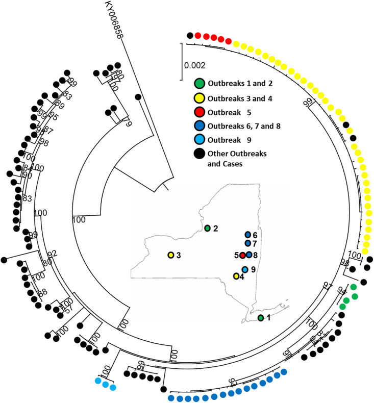
Since 2015, the United States has experienced a resurgence in the number of mumps cases and outbreaks in fully vaccinated populations. These outbreaks have occurred predominantly in close-quarter settings, such as camps, colleges, and detention centers. Phylogenetic analysis of 758 mumps-positive samples from outbreaks across the United States identified 743 (98%) as genotype G based on sequence analysis of the mumps small hydrophobic (SH) gene. Additionally, SH sequences in the genotype G samples showed almost no sequence diversity, with 675 (91%) of them having identical sequences or only one nucleotide difference. This uniformity of circulating genotype and strain created complications for epidemiologic investigations and necessitated the development of a system for rapidly generating mumps whole-genome sequences for more detailed analysis. In this study, we report a novel and streamlined assay for whole-genome sequencing (WGS) of mumps virus genotype G. The WGS procedure successfully generated 318 high-quality WGS sequences on nucleic acid from genotype G-positive respiratory samples collected during several mumps outbreaks in the United States between 2016 and 2019. Sequencing was performed by a rapid and highly sensitive custom Ion AmpliSeq mumps genotype G panel, with sample preparation performed on an Ion Chef and sequencing on an Ion S5. The WGS data generated by the AmpliSeq panel provided enhanced genomic resolution for epidemiological outbreak investigations. Translation and protein sequence analysis also identified several potentially important epitope changes in the circulating mumps genotype G strains compared to the Jeryl-Lynn strain (JL5) used in vaccines in the United States, which could explain the current level of vaccine escapes.

Keywords: AmpliSeq; genotyping; molecular epidemiology; mumps; vaccine; whole-genome sequencing.


Conflict of interest statement

P.B., H.C., D.M.L., and T.Y. declare no competing interests. K.S.G. receives research support from ThermoFisher for the evaluation of new assays for the detection and characterization of viruses. She also has a royalty-generating collaborative agreement with Zeptometrix.

Figures

FIG 1
FIG 1
Phylogenetic comparison of sequences from 63 mumps viruses from several outbreaks that occurred in New York State from 2016 to 2017. Samples from individual outbreaks are identified by specific colors, as indicated by the outbreak key. Sequence alignments were generated using the SH gene of the mumps genome (A), concatenated sequences of the SH, F, and HN genes (B), or the mumps whole-genome sequence (C). The evolutionary history was inferred by using the maximum likelihood method and Tamura-Nei model. The phylogeny was tested using 1,000 bootstrap replicates; values of >75 are indicated on the tree. The tree is drawn to scale, with branch lengths measured in the number of substitutions per site. Evolutionary analyses were conducted in MEGA X.
FIG 2
FIG 2
Mumps virus genotype G phylogenetic analysis with WGS. Maximum likelihood tree was constructed based on a nucleotide alignment of representative strains from five New York State outbreaks (color coded above). A map of New York State is included with the locations of the selected unique and related outbreaks. Additional individual samples and outbreak samples from across the United States are represented by black dots. Phylogenetic analysis was performed with MEGA X. Only bootstrap values of >50% are displayed.
FIG 3
FIG 3
Sequence alignment of the modified CDC mumps real-time RT-PCR assay in the NP gene of the consensus sequence of the 318 WGS genotype G genomes and additional genotypes A, C, G, H, I, J, and N. The modified CDC forward primer (MuN-687F) and probe (MuN-622P) are at the top of the alignment. Primer and probe degeneracy modifications in relation to the NP gene target consensus sequence are indicated above yellow boxes and boldfaced in gray. A third mutation located in the probe region of two samples from a 2017 outbreak in New Zealand that was not observed in the 318 WGS samples is indicated by an asterisk.
FIG 4
FIG 4
Density plot of age distribution for sporadic and outbreak-associated mumps cases. Data represent New York State, excluding New York City. Confirmed and probable cases were reported between 1 January 2016 and 1 June 2017. An outbreak is defined as three or more positive PCR results from a specific location, such as a university, county, or prison.
FIG 5
FIG 5
Donut chart of MMR status by percentage among reported mumps cases in New York State from January 2016 and June 2017.
FIG 6
FIG 6
In silico crystal structures modeling the epitope changes in the HN protein of the U.S. vaccine strain Jeryl Lynn 5 (A) compared to a representative genotype G sample (B) from 318 WGS samples. Blue arrows indicate potentially significant amino acid substitutions that were observed in four immunologically reactive regions labeled with their amino acid ranges and represented in magenta. The regions in yellow and white indicate the amino acids with alterations between Jeryl Lynn and the representative genotype G samples, pictured as stick models (A and B) or as spheres (C and D). Magnifications of the potentially significant Leu336Ser and Glu356Asp substitutions between Jeryl Lynn 5 (E) and the representative genotype G (F) epitope regions are represented in magenta, and sphere structures of amino acids are shown in white and yellow with amino acids adjacent to the changes in green. Structural modeling was performed using PyMOL 1.8.2.3, and mumps HN protein 5B2D at rcsb.org was used as a template (17).

References

    1. Hviid A, Rubin S, Muhlemann K. 2008. Mumps. Lancet 371:932–944. 10.1016/S0140-6736(08)60419-5. - DOI - PubMed
    1. Zamir CS, Schroeder H, Shoob H, Abramson N, Zentner G. 2015. Characteristics of a large mumps outbreak: clinical severity, complications and association with vaccination status of mumps outbreak cases. Hum Vaccin Immunother 11:1413–1417. 10.1080/21645515.2015.1021522. - DOI - PMC - PubMed
    1. Jin L, Rima B, Brown D, Orvell C, Tecle T, Afzal M, Uchida K, Nakayama T, Song JW, Kang C, Rota PA, Xu W, Featherstone D. 2005. Proposal for genetic characterisation of wild-type mumps strains: preliminary standardisation of the nomenclature. Arch Virol 150:1903–1909. 10.1007/s00705-005-0563-4. - DOI - PubMed
    1. Jin L, Orvell C, Myers R, Rota PA, Nakayama T, Forcic D, Hiebert J, Brown KE. 2015. Genomic diversity of mumps virus and global distribution of the 12 genotypes. Rev Med Virol 25:85–101. 10.1002/rmv.1819. - DOI - PubMed
    1. Centers for Disease Control and Prevention. 1978. Mumps surveillance-United States. MMWR Morb Mortal Wkly Rep 40:379–381.

Publication types