. 2021 Nov 10;19(11):e3001447.
doi: 10.1371/journal.pbio.3001447. eCollection 2021 Nov.

p38γ and p38δ regulate postnatal cardiac metabolism through glycogen synthase 1

Affiliations

p38γ and p38δ regulate postnatal cardiac metabolism through glycogen synthase 1

Ayelén M Santamans et al. PLoS Biol. .

Abstract

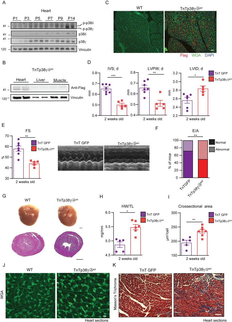
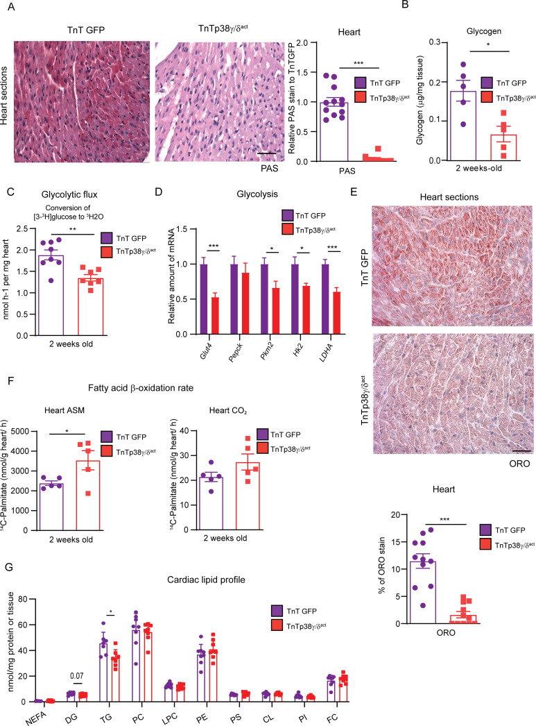
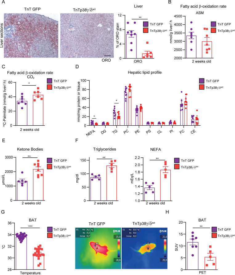
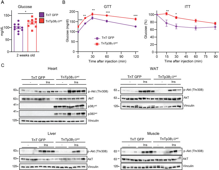
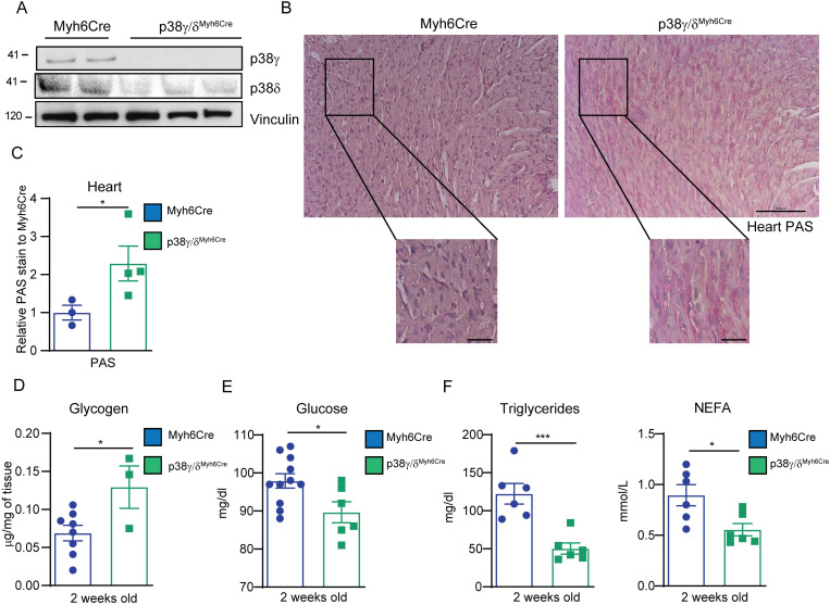
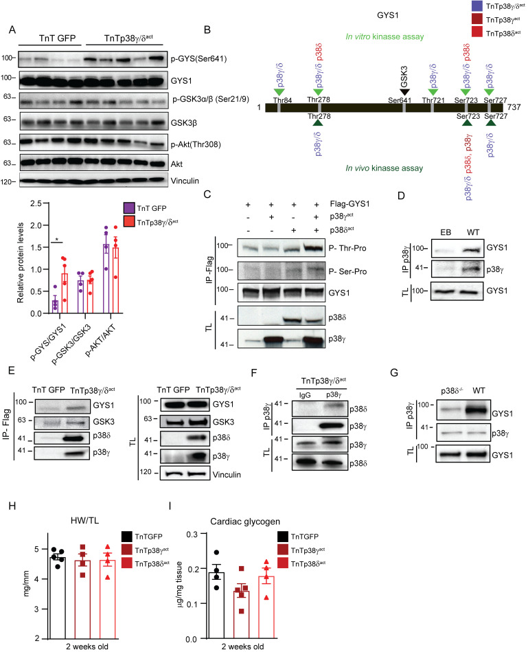
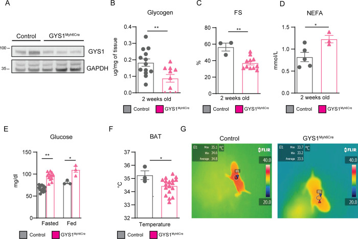
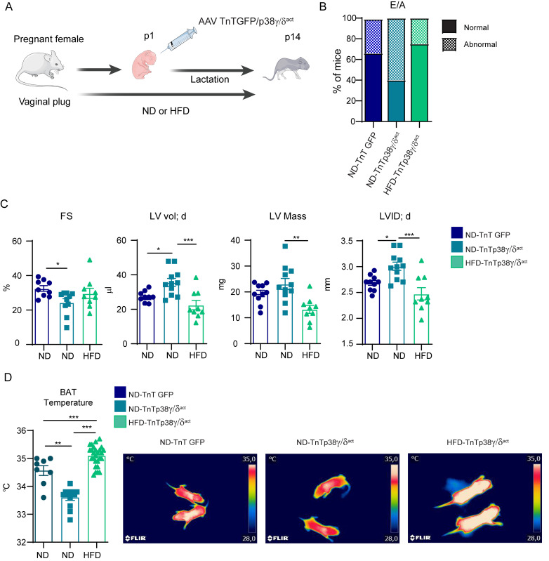
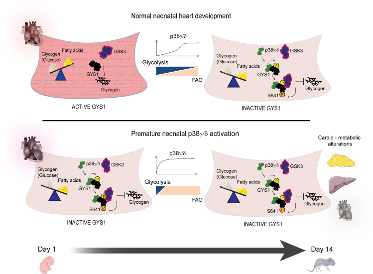
During the first weeks of postnatal heart development, cardiomyocytes undergo a major adaptive metabolic shift from glycolytic energy production to fatty acid oxidation. This metabolic change is contemporaneous to the up-regulation and activation of the p38γ and p38δ stress-activated protein kinases in the heart. We demonstrate that p38γ/δ contribute to the early postnatal cardiac metabolic switch through inhibitory phosphorylation of glycogen synthase 1 (GYS1) and glycogen metabolism inactivation. Premature induction of p38γ/δ activation in cardiomyocytes of newborn mice results in an early GYS1 phosphorylation and inhibition of cardiac glycogen production, triggering an early metabolic shift that induces a deficit in cardiomyocyte fuel supply, leading to whole-body metabolic deregulation and maladaptive cardiac pathogenesis. Notably, the adverse effects of forced premature cardiac p38γ/δ activation in neonate mice are prevented by maternal diet supplementation of fatty acids during pregnancy and lactation. These results suggest that diet interventions have a potential for treating human cardiac genetic diseases that affect heart metabolism.


Conflict of interest statement

The authors have declared that no competing interests exist.

Figures

Fig 1
Fig 1. Cardiomyocyte-specific postnatal overexpression of p38γ/δact causes cardiac eccentric hypertrophy and heart dysfunction.
(A) Immunoblot analysis of endogenous p38γ/δ in heart extracts from WT (non-injected) mice at PD 1, 3, 5, 7, 9, and 14. (B) Immunoblot using anti-FLAG antibody showing p38γ/δact specific cardiac overexpression at PD14 after AAV-cTnT-p38γ/δact injection (at PD1), in heart, liver, and muscle extracts. (C) Immunofluorescence of FLAG-p38γ/δact (red), WGA (green), and DAPI (blue) on heart sections from WT or TnTp38γ/δact mice. Scale bar: 200 μm. (D–K) Analyses of hearts at PD14 of AAV-cTnT-GFP-Luc (TnTGFP; control mice) or TnTp38γ/δact mice (with AAV injection at PD1), showing the following: (D) echocardiography measurements. (E) FS and M-mode short-axis echocardiography traces; (F) percentage of mice at PD14 with normal or abnormal mitral valve flow (E/A) as an indicator of diastolic dysfunction; (G) images of whole heart (scale bar: 1 mm) and H&E staining of transverse heart sections (scale bar: 1 mm); (H) HWTL ratio; (I, J) chart of FITC-WGA staining of hearts (green), with cardiomyocyte cross-sectional area quantification (scale bar: 50 μm); (K) Masson’s trichrome staining images from heart sections (the respective fibrosis quantification is shown in S1B Fig). Scale bar: 200 μm. Data are mean ± SEM (n = 5–8). *p < 0.05; **p < 0.01; ***p < 0.001 by two-tailed Student t test. Raw data are given in S14 Fig. AAV, adeno-associated virus; FS, fractional shortening; HWTL, heart weight-to-tibia length; H&E, hematoxylin and eosin; IVS; d, interventricular septum thickness in diastole; LVID; d, left ventricular internal diameter in diastole; LVPW; d, left ventricle posterior wall thickness in diastole; PD, postnatal day; WT, wild-type.
Fig 2
Fig 2. Cardiac-specific p38γ/δact overexpression decreases cardiac glycogen storage and lipid accumulation.
Mice were IV injected at PD1 with AAV-cTnT-GFP-Luc (TnTGFP) or AAV-cTnT-p38γ/δact (TnTp38γ/δact) and killed at PD14. (A) PAS staining in heart sections of TnTp38γ/δact or TnTGFP mice (left) and quantification (right). Scale bar: 200 μm. (B) Cardiac glycogen quantification. (C) Conversion of [3-3H] glucose into 3H2O, reflecting the glycolytic rate in hearts from TnTGFP and TnTp38γ/δact mice. (D) qRT-PCR analysis of glycolytic enzymes expression in cardiac tissue. (E) ORO staining in heart sections, showing representative images. Quantification chart below. Scale bar: 50 μm. (F) Cardiac fatty acid oxidation rate of 14C-palmitate determined by the production of CO2 and ASMs. (G) Cardiac lipid profile. All lipid amounts were normalized by mg of protein except for NEFAs, which were relativized by mg of tissue. Data are mean ± SEM. (n = 5–12). *p < 0.05; **p < 0.01; ***p < 0.001 by Student t test. Raw data are given in S14 Fig. ASM, acid-soluble metabolite; CL, cardiolipin; DG, diglycerides; FC, free cholesterol; LPC, lysophosphatidylcholine; NEFA, non-esterified fatty acid; ORO, oil red O; PAS, periodic acid–Schiff; PC, phosphatidylcholine; PE, phosphatidylethanolamine; PI, phosphatidylinositol; PS, phosphatidylserine; qRT-PCR, real-time quantitative PCR; TG, triglyceride.
Fig 3
Fig 3. Cardiac-specific p38γ/δact overexpression has whole-body metabolic consequences.
Mice were IV injected at PD1 with AAV-cTnT-GFP-Luc (TnTGFP) or AAV-cTnT-p38γ/δact (TnTp38γ/δact); after metabolic tests were performed at PD14, mice were killed. (A) ORO staining (left) and quantification (right) of liver sections. Scale bar: 50 μm. (B, C) Hepatic fatty acid oxidation rate of 14C-palmitate, determined by the production of ASMs and CO2. (D) Hepatic lipid profile. All lipid amounts were normalized by mg of protein except for NEFAs, which were relativized to mg of tissue. (E) Plasma ketone bodies. (F) Plasma TG and NEFA. (G) Mice BAT temperatures, with its representative thermographic images. (H) BAT glucose uptake measured by PET-CT. Regions of interest were delimited to the BAT area to obtain the mean SUV. Data are mean ± SEM (n = 7–10). *p < 0.05, **p < 0.01; ***p < 0.001, by Student t test. Raw data are given in S14 Fig. ASM, acid-soluble metabolite; BAT, brown adipose tissue; CE, cholesteryl ester; CL, cardiolipin; DG, diglyceride; FC, free cholesterol; LPC, lysophosphatidylcholine; NEFA, non-esterified fatty acid; ORO, oil red O; PC, phosphatidylcholine; PE, phosphatidylethanolamine; PET-CT, positron emission tomography–computed tomography; PI, phosphatidylinositol; PS, phosphatidylserine; SUV, standard uptake value; TG, triglyceride.
Fig 4
Fig 4. Cardiac-specific p38γ/δact overexpression leads to glucose intolerance and insulin resistance.
Mice were treated as for Fig 3. (A) Blood glucose levels. (B) GTT and ITT. (C) Immunoblot analysis of insulin-stimulated Akt phosphorylation (at Thr308) in heart, WAT, liver, and skeletal muscle. Mice at PD14 were killed 15 min after IP insulin injection. Data are mean ± SEM (n = 7–10). *p < 0.05; **p < 0.01; ***p < 0.001 by Student t test or two-way ANOVA coupled to Tukey posttest. Raw data are given in S14 Fig. GTT, glucose tolerance test; ITT, insulin tolerance test; WAT, white adipose tissue.
Fig 5
Fig 5. Postnatal cardiac-specific p38γ/δact overexpression leads to heart defects that persist throughout life with no metabolic consequences in adulthood.
Mice were IV injected at PD1 with AAV-cTnT-GFP-Luc (TnTGFP) or AAV-cTnT-p38γ/δact (TnTp38γ/δact), analyzed for phenotype progression over 12 weeks, and then killed. (A) Immunoblot analysis of exogenous active (p38γact) and endogenous p38γ in hearts extracts of 12-week-old mice. (B, C) Masson’s trichrome staining in heart sections from 12-weeks-old mice (B), and quantification (C). Scale bar: 200 μm. (D) Left ventricular FS progression from weeks 2 to 12. (E) Plasma glucose. (F) GTT and ITT. (G) Plasma NEFA. (H) Plasma triglycerides. (I) Representative PAS staining of heart sections with its respective quantification relative to TnTGFP mice. Scale bar: 200 μm. Data are mean ± SEM (n = 4–8). *p < 0.05; **p < 0.01; ***p < 0.001 by two-way ANOVA coupled to Tukey posttest or Student t test. Raw data are given in S14 Fig. FS, fractional shortening; GTT, glucose tolerance test; ITT, insulin tolerance test; NEFA, non-esterified fatty acid; PAS, periodic acid–Schiff.
Fig 6
Fig 6. Early postnatal cardiac-specific p38γ and p38δ deletion increases cardiac glycogen storage and affects whole body metabolism.
Mice were IP injected with tamoxifen (p38γ/δ Myh6-Cre) or vehicle (Myh6-Cre; control mice) at PD1, PD2, and PD3, and killed at PD14. (A) Immunoblot analysis of p38γ and p38δ in heart extracts. (B, C) Representative images of PAS staining of heart sections (B) and its quantification, normalized to Myh6-Cre (C). Scale bar: 100 μm and 25 μm (amplification). (D) Glycogen quantification. (E) Blood glucose levels. (F) Plasma triglycerides and NEFA. Data are mean ± SEM (n = 4–6). *p < 0.05; ***p < 0.001 by Student t test. Raw data are given in S14 Fig. NEFA, non-esterified fatty acid; PAS, periodic acid–Schiff.
Fig 7
Fig 7. p38γ/δ cooperatively interact with GYS1 and GSK3 to promote GYS1 phosphorylation at its canonical site (Ser641).
(A) Immunoblot analysis of the Akt-GSK3–GYS axis in cardiac homogenates from AAV-cTnT-GFP-Luc (TnTGFP) or AAV-cTnT-p38γ/δact (TnTp38γ/δact) mice killed at PD14 with its respective quantification (lower panel). (B) Scheme showing GYS1 sites phosphorylated by p38γ, p38δ, or both, in an in vitro kinase assay (top, light green) or an in vivo kinase assay in HEK-293 cells (bottom, dark green). The GYS1 canonical site for GSK3 phosphorylation is Ser641 (shown in bold). Data are representative of at least 3 independent experiments (biological replicates). (C) In vivo phosphorylation of GYS1 in HEK-293 cells that had been transfected with Flag-GYS1 alone or together with p38γact or p38δ act, or both. Phosphorylation of transfected GYS1 was evaluated in the Flag-immunoprecipitate with phospho-MAPK substrates for Ser or Thr. TL before immunoprecipitation is shown as control. (D) Immunoprecipitation and immunoblot analysis of GYS1 and endogenous p38γ association in WT mice. (E) Immunoblot analysis of GYS1 and GSK3 in Flag-p38γ/δact immunoprecipitates from heart lysates, to detect interactions between these proteins and the exogenous p38γ/δact. (F) Immunoprecipitation and immunoblot analysis of the association between p38δ and p38γ in TnTp38γ/δact mice. (G) Immunoprecipitation/immunoblot analysis of the interactions between GYS1 and endogenous p38γ in WT or p38δ−/− mice. (H, I) Analysis of hearts from mice that were IV injected at PD1 with AAV-cTnT-GFP-Luc (TnTGFP), AAV-cTnT-p38γact (TnTp38γact), or AAV-cTnT-p38δact (TnTp38δact) and killed at PD14, showing (H) HWTL ratio and (I) cardiac glycogen content. Data are mean ± SEM (n = 10). One-way ANOVA coupled to Tukey posttest or Student t test. Raw data are given in S14 Fig. EB, empty bead; GSK3, glycogen synthase kinase-3; GYS1, glycogen synthase 1; HWTL, heart weight to tibia length; TL, total lysate; WT, wild-type.
Fig 8
Fig 8. Postnatal GYS1 deletion leads to whole-body metabolic alterations and cardiac dysfunction.
Gys1Myh6Cre or Myh6-Cre (control) mice were IP injected with 62.5 mg/kg tamoxifen at PD1, PD2, and PD3, and killed at PD14. (A) Immunoblot showing partial GYS1 deletion in heart extracts. (B) Heart glycogen content. (C) Echocardiography-measured FS. (D) NEFA plasma levels. (E) Blood plasma glucose in fed or fasted (e.g., food deprived for 4 h) conditions. (F, G) BAT temperature chart and representative thermographic images mice at PD14. Data are mean ± SEM (n = 3–17). *p < 0.05; **p < 0.01; ***p < 0.001 by Student t test or two-way ANOVA coupled to Tukey posttest. Raw data are given in S14 Fig. BAT, brown adipose tissue; FS, fractional shortening; GYS1, glycogen synthase 1; NEFA, non-esterified fatty acid.
Fig 9
Fig 9. Maternal HFD feeding suppresses cardiac dysfunction in pups overexpressing p38γ/δact.
(A) Schematic protocol: CD1 females were crossed; after pregnancy confirmation by vaginal plug appearance, they were fed a HFD for the entire experiment (e.g., pregnancy and lactation). Neonates were IV injected at PD1 with AAV-cTnT-GFP-Luc (TnTGFP) or AAV-cTnT-p38γ/δact (TnTp38γ/δact); during their lactation, mother remained on the same diet (e.g., ND or HFD) as during pregnancy. Pups were killed at PD14. (B) Percentage of mice at PD14 with normal or abnormal mitral valve flow (E/A) as an indicator of diastolic dysfunction. (C) Echocardiography measured parameters. (D) BAT temperature chart and representative thermographic images from 2-week-old mice. Data are mean ± SEM (n = 9 or 10). *p < 0.05, **p < 0.01, ***p < 0.001 by one-way ANOVA coupled to Tukey posttest or chi-squared test. Raw data are given in S14 Fig. The figure was prepared using Servier Medical Art (https://smart.servier.com/). BAT, brown adipose tissue; FS, fractional shortening; HFD, high-fat diet; LVID; d, left ventricular internal diameter in diastole; LV mass, left vetricular mass; LVvol; d, left ventricular volume in diastole; ND, normal diet.
Fig 10
Fig 10. Early cardiac p38γ/δ expression inactivates GYS1 affecting cardiac and whole-body metabolism, but the effects can be mitigated by metabolic intervention.
Schematic overview of our findings At early postnatal development, p38γ/δ are not present in heart and in consequence, they did not facilitate GYS phosphorylation and inactivation by GSK3 allowing glycogen storage. Premature postnatal cardiac-specific p38γ/δ overexpression triggers a premature metabolic switch to fatty acid oxidation, with whole-body metabolic alterations, including insulin resistance, glucose intolerance, altered hepatic lipid metabolism, and impaired thermogenesis, as well as permanent cardiac defects. The 2 kinases, p38γ/δ, work collaboratively to control cardiac glycogen storage by regulating GYS1’s interaction with its inhibitory kinase GSK3. Maternal metabolic intervention by HFD feeding during pregnancy and lactation mitigated the cardiac dysfunction and impaired thermogenesis in offspring, setting a precedent for treatment of neonatal cardiometabolic genetic diseases. The figure was prepared using Servier Medical Art (https://smart.servier.com/). FAO, fatty acid oxidation; GSK3, glycogen synthase kinase-3; GYS1, glycogen synthase 1; HFD, high-fat diet.

References

    1. Ellen Kreipke R, Wang Y, Miklas JW, Mathieu J, Ruohola-Baker H. Metabolic remodeling in early development and cardiomyocyte maturation. Semin Cell Dev Biol. 2016;52:84–92. Epub 2016 Feb 26. doi: 10.1016/j.semcdb.2016.02.004 ; PubMed Central PMCID: PMC4820352. - DOI - PMC - PubMed
    1. Lopaschuk GD, Jaswal JS. Energy metabolic phenotype of the cardiomyocyte during development, differentiation, and postnatal maturation. J Cardiovasc Pharmacol. 2010;56(2):130–40. Epub 2010 May 28. doi: 10.1097/FJC.0b013e3181e74a14 . - DOI - PubMed
    1. Grynberg A, Demaison L. Fatty acid oxidation in the heart. J Cardiovasc Pharmacol. 1996;28(Suppl 1):S11–7. Epub 1996 Jan 1. doi: 10.1097/00005344-199600003-00003 . - DOI - PubMed
    1. Pederson BA, Chen H, Schroeder JM, Shou W, DePaoli-Roach AA, Roach PJ. Abnormal cardiac development in the absence of heart glycogen. Mol Cell Biol. 2004;24(16):7179–87. Epub 2004 Jul 30. doi: 10.1128/MCB.24.16.7179-7187.2004 ; PubMed Central PMCID: PMC479719. - DOI - PMC - PubMed
    1. Chung S, Dzeja PP, Faustino RS, Perez-Terzic C, Behfar A, Terzic A. Mitochondrial oxidative metabolism is required for the cardiac differentiation of stem cells. Nat Clin Pract Cardiovasc Med. 2007;4(Suppl 1):S60–7. Epub 2007 Jan 19. doi: 10.1038/ncpcardio0766 ; PubMed Central PMCID: PMC3232050. - DOI - PMC - PubMed

Publication types

MeSH terms