. 2021 Nov 10;17(11):e1009957.
doi: 10.1371/journal.ppat.1009957. eCollection 2021 Nov.

A single amino acid polymorphism in a conserved effector of the multihost blast fungus pathogen expands host-target binding spectrum

Affiliations

A single amino acid polymorphism in a conserved effector of the multihost blast fungus pathogen expands host-target binding spectrum

Adam R Bentham et al. PLoS Pathog. .

Abstract

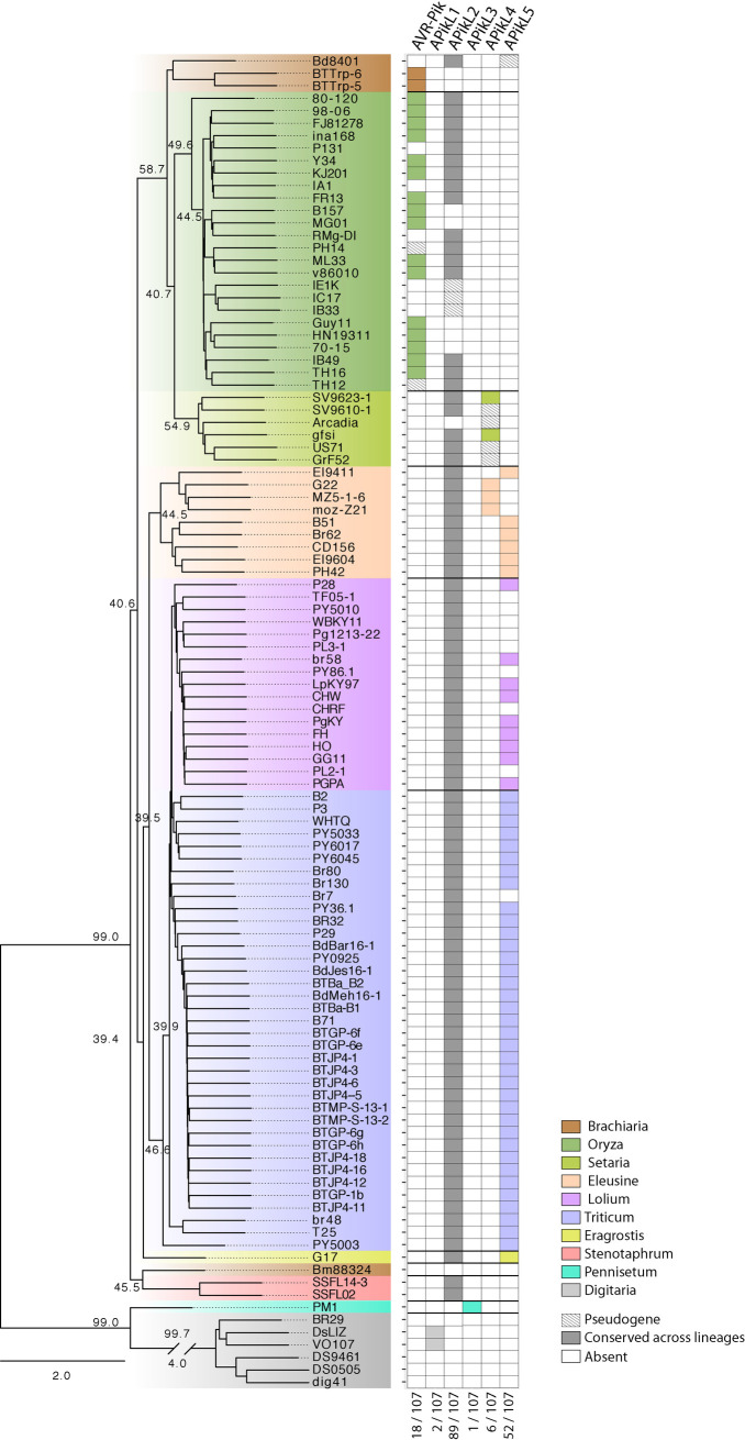
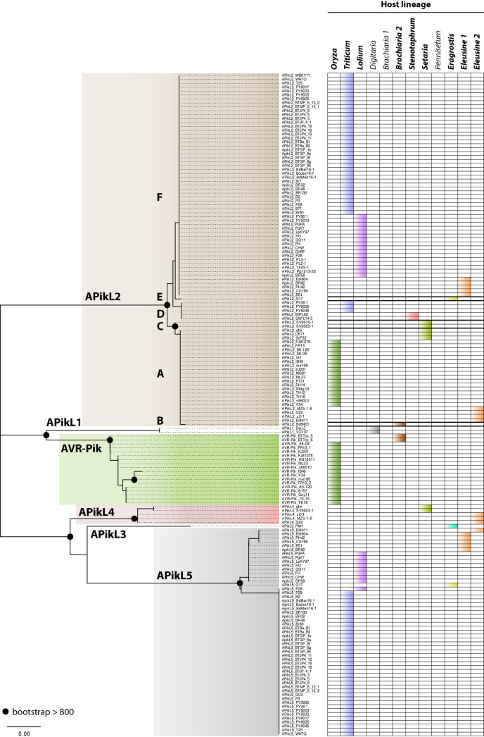
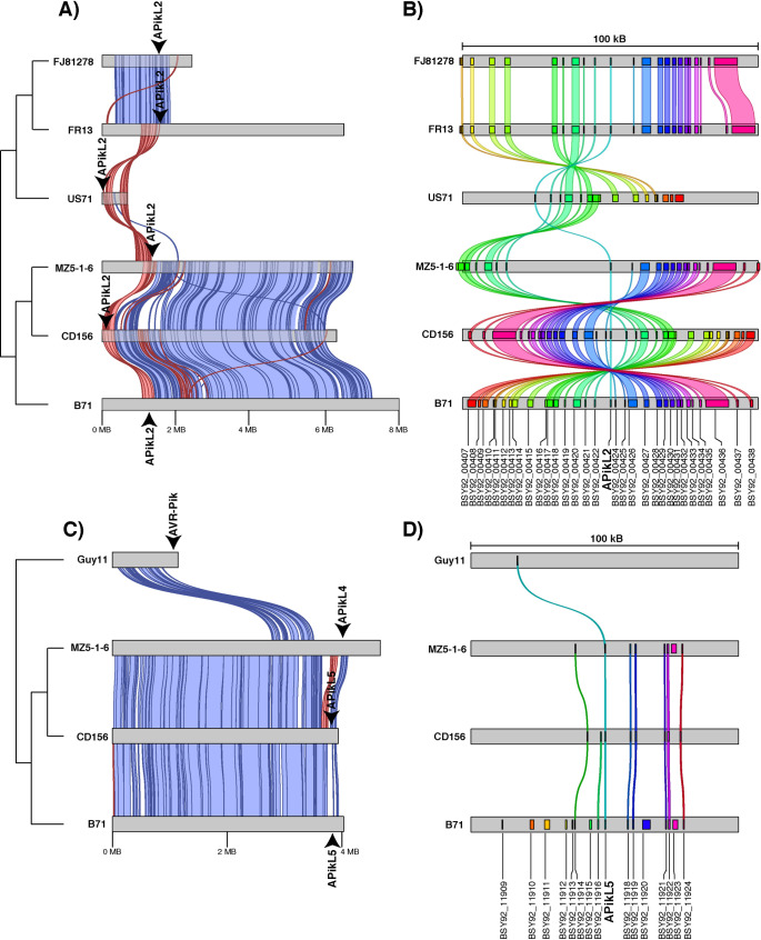
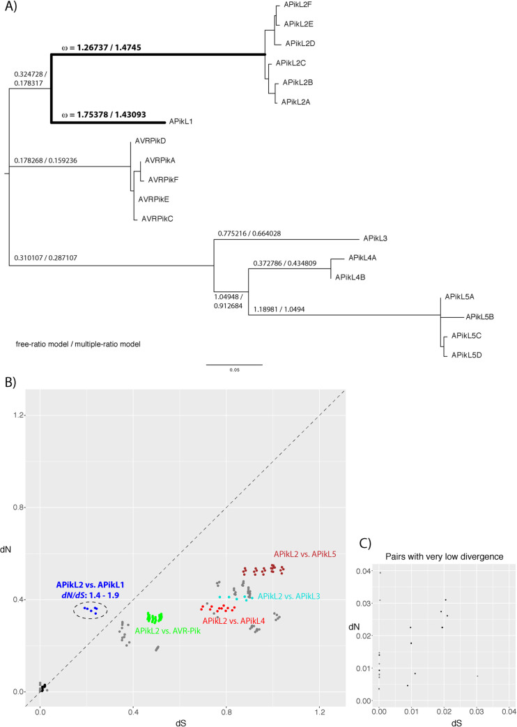
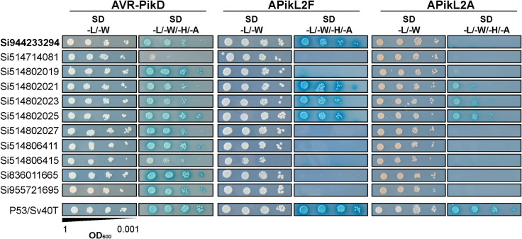
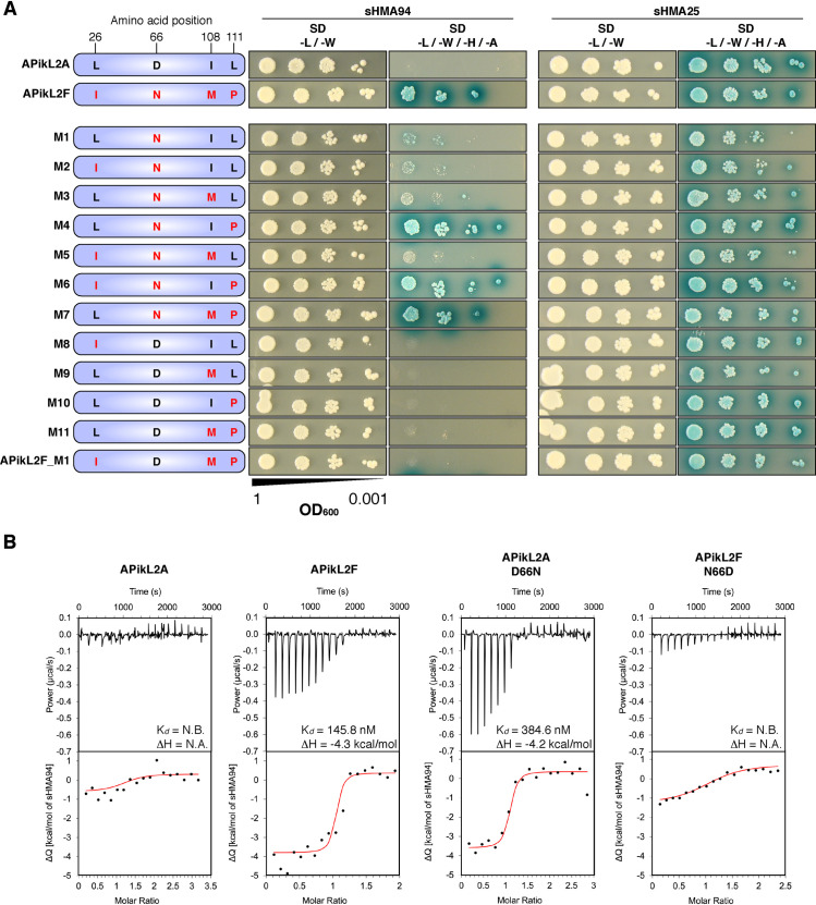
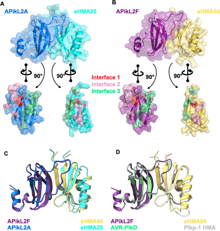
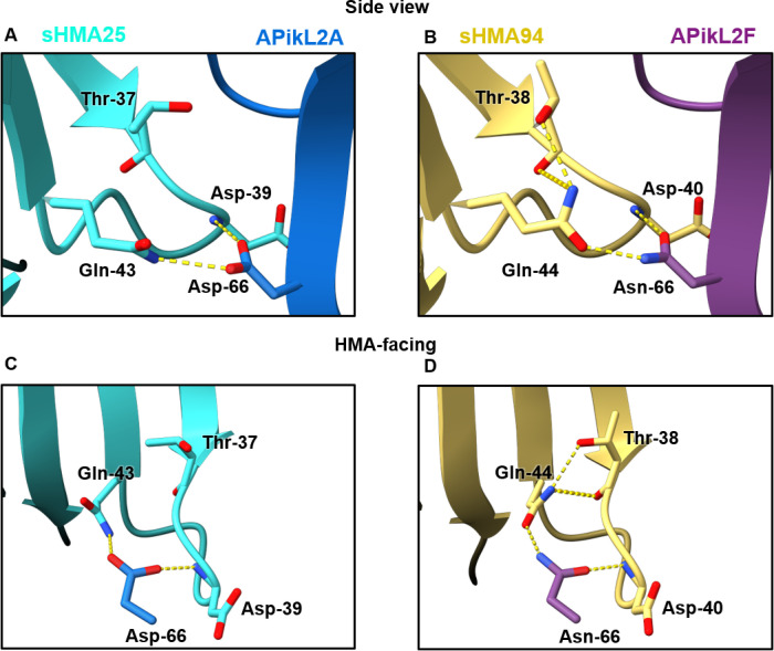
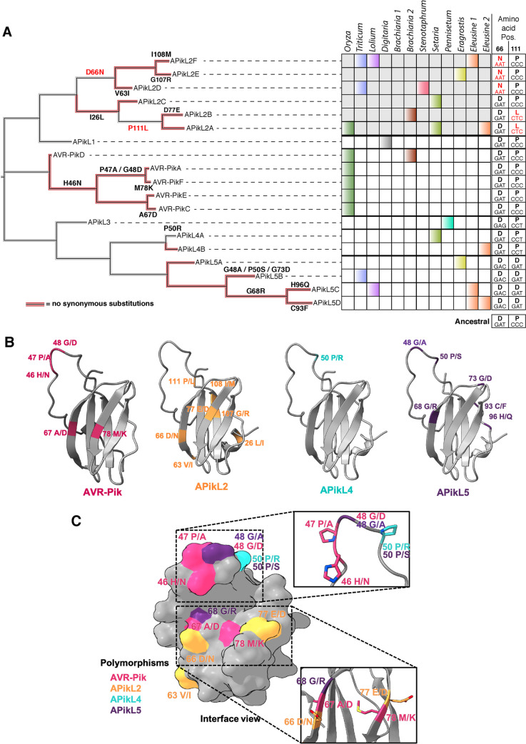
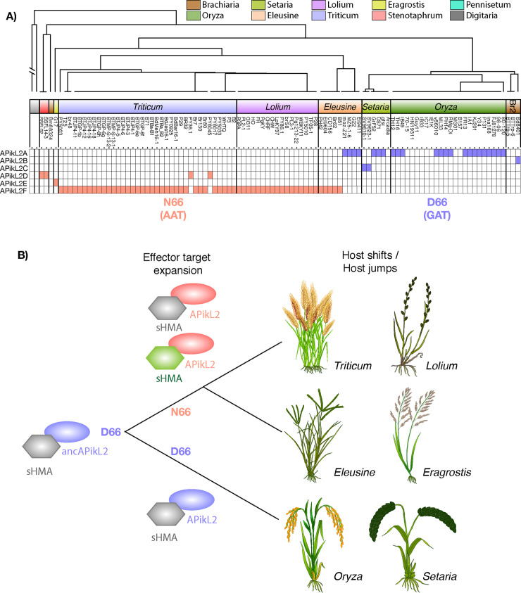
Accelerated gene evolution is a hallmark of pathogen adaptation and specialization following host-jumps. However, the molecular processes associated with adaptive evolution between host-specific lineages of a multihost plant pathogen remain poorly understood. In the blast fungus Magnaporthe oryzae (Syn. Pyricularia oryzae), host specialization on different grass hosts is generally associated with dynamic patterns of gain and loss of virulence effector genes that tend to define the distinct genetic lineages of this pathogen. Here, we unravelled the biochemical and structural basis of adaptive evolution of APikL2, an exceptionally conserved paralog of the well-studied rice-lineage specific effector AVR-Pik. Whereas AVR-Pik and other members of the six-gene AVR-Pik family show specific patterns of presence/absence polymorphisms between grass-specific lineages of M. oryzae, APikL2 stands out by being ubiquitously present in all blast fungus lineages from 13 different host species. Using biochemical, biophysical and structural biology methods, we show that a single aspartate to asparagine polymorphism expands the binding spectrum of APikL2 to host proteins of the heavy-metal associated (HMA) domain family. This mutation maps to one of the APikL2-HMA binding interfaces and contributes to an altered hydrogen-bonding network. By combining phylogenetic ancestral reconstruction with an analysis of the structural consequences of allelic diversification, we revealed a common mechanism of effector specialization in the AVR-Pik/APikL2 family that involves two major HMA-binding interfaces. Together, our findings provide a detailed molecular evolution and structural biology framework for diversification and adaptation of a fungal pathogen effector family following host-jumps.


Conflict of interest statement

The authors have declared that no competing interests exist.

Figures

Fig 1
Fig 1. APikL2 is conserved across different host-specific lineages of M. oryzae.
Presence/absence analysis of APikL family members shows conservation of APikL2 across all host-specific lineages of M. oryzae. All other family members show presence/absence polymorphisms that correlate with the species phylogeny and host-specificity. Left: ASTRAL multispecies coalescence tree derived from 1920 maximum-likelihood trees of all conserved single copy orthologs. Numbers at selected main branches represent the q1 local quartet tree support of individual genealogies. Right: Presence of APikL family members in each isolate. Colors indicate the host-specific lineages. The numbers at the bottom show the number of isolates that contain a certain effector.
Fig 2
Fig 2. APikL2 sequence diversification is associated with host-specific lineages.
Left: APikL family maximum likelihood tree based on amino acid sequences of all APikL proteins from 107 M. oryzae genomes. Colors indicate APikL family members where allelic diversification was observed in our dataset. Black circles indicate a bootstrap support >800 of the major nodes of allelic variants. Right: Presence of APikL family members in various host-specific lineages. Colors indicate host lineages as shown in Fig 1. Host-lineages in bold indicate presence of APikL2.
Fig 3
Fig 3. The APikL2 locus is conserved across diverse genetic lineages of M. oryzae.
Synteny analysis of APikL loci in six isolates from five genetic M. oryzae lineages. A) Pairwise alignments of contigs containing the APikL2 gene. Forward and reverse alignments are shown in blue and red, respectively. Arrowheads indicate the position of the APikL2 genes in the assemblies. B) Conservation of genes in a 100 kB region surrounding the APikL2 locus. Colored links indicate homologous genes. C) Pairwise alignments of contigs containing diverse APikL family members on chromosome 7. Forward and reverse alignments are shown in blue and red, respectively. Arrowheads indicate the position of APikL family genes in the assemblies. Genetic relationship of the isolates is shown schematically on the left. D) Conservation of genes in a 100 kB region surrounding the loci of APikL family members on chromosome 7. Colored links indicate homologous genes.
Fig 4
Fig 4. APikL2 displays patterns of positive selection.
A) Maximum-likelihood tree of all APikL family members based on nucleotide sequences. Numbers on branches of the tree represent ω (dN/dS ratio of non-synonymous and synonymous substitutions) as calculated according to the free-ratio and multiple-ratio model implemented in PAML. Branches with high ω leading to APikL2 and APikL1 are highlighted in bold. B) Comparison of pairwise dN/dS ratios between APikL family members. Colors indicate pairwise comparisons between APikL2 and other APikL family members. dN/dS ratios between other APikL family members are shown in grey. Pairwise comparisons between APikL2 variants are shown in black. The dotted line indicates dN/dS = 1. C) Enlarged view of pairwise comparisons in B with low divergence.
Fig 5
Fig 5. APikL2 variants interact differentially with the HMA-domain of Setaria italica protein sHMA94.
Subset of the pairwise yeast two-hybrid screen to identify interaction partners of AVR-Pik and the APikL2 variants APikL2A and APikL2F from the Oryza- and Triticum-infecting lineages of M. oryzae. All positive interactions between S. italica HMA-domains with the APikL family members AVR-Pik and APikL2 are shown. Left: gene identifiers of HMA-domain containing proteins. SD -L/-W: Synthetic double dropout medium lacking the amino acids leucin and tryptophan; SD -L/-W/-H/-A: Synthetic quadruple dropout medium lacking the amino acids leucine, tryptophan, histidine, and adenine.
Fig 6
Fig 6. Two polymorphic sites determine binding specificity of APikL2 variants to sHMA94.
A) Pairwise yeast two-hybrid assays between sHMA94 and APikL2A, APikL2F, and 12 APikL2 mutants derived from combinations of the four polymorphic residues. The two naturally occurring variants, APikL2A and APikL2F, were used as positive and negative controls. Mutations in each variant are shown schematically (left). sHMA25 was used as a positive control for both APikL2 variants. SD -L/-W: Synthetic double dropout medium lacking the amino acids leucine and tryptophan; SD -L/-W/-H/-A: Synthetic quadruple dropout medium lacking the amino acids leucine, tryptophan, histidine, and adenine. B) Asn-66 is essential for binding of APikL2 to sHMA94 in vitro. In vitro binding affinities for sHMA94 and APikL2 proteins were assessed by ITC. The top panels show heat differences upon injection of the effector into the cell containing sHMA94. The lower panel shows integrated heats of injection (•) and the best fit (red line) to a single site binding model calculated using AFFINImeter ITC analysis software. APikL2F, but not APikL2A, binds sHMA94. The APikL2F Asn-66 to Asp-66 polymorphism disrupts sHMA94 binding. Conversely, the APikL2A Asp-66 to Asn-66 polymorphism facilitates binding of sHMA94, with affinity similar to APikL2F.
Fig 7
Fig 7. Crystal structures of APikL2 in complex with HMA domains reveals a conserved binding interface.
A) Cartoon and surface representation of the APikL2A/sHMA25 complex, with APikL2A coloured blue and sHMA25 coloured cyan. B) Cartoon and surface representation of the APikL2F/sHMA94 complex, with APikL2F coloured purple and sHMA94 coloured yellow. The effector was separated from the complex using PyMol and rotated 90° relative to the complex to give an interior view of the interaction interface. The residues that form the interfaces one, two, and three (as defined by [7]), between the effector and HMA are coloured dark blue, pink, and magenta, respectively. C) Superimposition of the APikL2F/sHMA94 (purple and yellow) and APikL2A/sHMA25 (blue and cyan) complexes performed with PyMol. D) Structural alignment of APikL2F/sHMA94 (purple and yellow) with AVR-PikD/Pikp-1 HMA (green and grey; PDB ID: 6G10).
Fig 8
Fig 8. The aspartate/asparagine polymorphism at position 66 in APikL2 results in an altered hydrogen bonding network at the sHMA interface.
A, C) The carboxylic side chain of Asp-66 from APikL2A forms two hydrogen bonds with sHMA25 through the NE2 atom of Gln-43 and the backbone nitrogen of Asp-39. B, D) The amide side chain of Asn-66 from APikL2F forms two hydrogen bonds with sHMA94 through the OE1 atom of Gln-44 and the backbone amide of Asp-40. The interaction with Gln-44 via the OE1 atom in the AVR-Pik2F/ sHMA structure flips Gln-44 respective to the Gln-43 residue of the APikL2A/sHMA25 structure. This reorientation of Gln-44 in sHMA94 allows the NE2 atom of Gln-44 to form intramolecular hydrogens bond with the OG1 atom and backbone carbonyl of Thr-38.
Fig 9
Fig 9. Adaptive mutations shape the binding interfaces of APikL family effectors.
A) Asn-66 is a recently derived polymorphism in APikL2 effectors. Left: Reconstruction of the evolutionary history of the APikL family. Multiple non-synonymous mutations contribute to allelic diversification, largely in absence of synonymous mutations (red branches). Right: APikL effector presence/absence across multiple host-specific lineages. Colors correspond to host lineages as in Fig 1. Amino acids and codons of positions 66 and 111 across the APikL family are shown on the right. Acquired mutations are highlighted in red. The ancestral states are shown at the bottom. B) Mapping of non-synonymous changes that contribute to allelic diversification to the structures of APikL family members AVR-Pik, APikL2, APikL4, and APikL5. Divergent amino acids of each effector are highlighted. C) Allelic diversification reveals two major hotspots across the APikL family. Surface representation of divergent sites across the APikL family combining all residues shown in B. Most polymorphic residues reside in the HMA-binding interfaces 1 (bottom) and 2 (top).
Fig 10
Fig 10. The Asp-66-Asn polymorphism predates the emergence of host-adapted lineages of M. oryzae.
A) Distribution of the critical Asp-66 to Asn-66 mutation in M. oryzae populations. Schematic illustration of host-specific lineages of M. oryzae and annotation of the Asp-66/Asn-66 polymorphism. Blue: Asp-66; Red: Asn-66. The host genera of the different M. oryzae lineages are shown. B) Model illustrating the emergence of the Asn-66 polymorphism prior to the differentiation of host specialized lineages. Blue: APikL2 variants carrying Asp-66; Red: APikL2 variants carrying Asn-66; Grey: sHMA target protein interacting with both APikL2 variants; Green: New sHMA target protein due to Asn-66 polymorphism.

Similar articles

Cited by

References

    1. Dong S, Stam R, Cano LM, Song J, Sklenar J, Yoshida K, et al.. Effector Specialization in a Lineage of the Irish Potato Famine Pathogen. Science. 2014. Jan 31;343(6170):552. doi: 10.1126/science.1246300 - DOI - PubMed
    1. Kim K-T, Ko J, Song H, Choi G, Kim H, Jeon J, et al.. Evolution of the Genes Encoding Effector Candidates Within Multiple Pathotypes of Magnaporthe oryzae. Front Microbiol. 2019;10:2575. doi: 10.3389/fmicb.2019.02575 - DOI - PMC - PubMed
    1. Kourelis J, Malik S, Mattinson O, Krauter S, Kahlon PS, Paulus JK, et al.. Evolution of a guarded decoy protease and its receptor in solanaceous plants. Nat Commun. 2020. Sep 2;11(1):4393. doi: 10.1038/s41467-020-18069-5 - DOI - PMC - PubMed
    1. Upson JL, Zess EK, Białas A, Wu C-H, Kamoun S. The coming of age of EvoMPMI: evolutionary molecular plant-microbe interactions across multiple timescales. Curr Opin Plant Biol. 2018. Aug;44:108–16. doi: 10.1016/j.pbi.2018.03.003 - DOI - PubMed
    1. Dong S, Raffaele S, Kamoun S. The two-speed genomes of filamentous pathogens: waltz with plants. Curr Opin Genet Dev. 2015. Dec;35:57–65. doi: 10.1016/j.gde.2015.09.001 - DOI - PubMed

Publication types