Case Reports
. 2021 Nov 10;21(1):423.
doi: 10.1186/s12876-021-02009-8.

Pulmonary tumor thrombotic microangiopathy in occult early gastric cancer that was undetectable on upper endoscopy: a case report and review of similar cases

Affiliations
Case Reports

Pulmonary tumor thrombotic microangiopathy in occult early gastric cancer that was undetectable on upper endoscopy: a case report and review of similar cases

Naoki Kawakami et al. BMC Gastroenterol. .

Abstract

Background: Pulmonary tumor thrombotic microangiopathy (PTTM), a rare manifestation of metastatic cancer with poor prognosis, is characterized by subacute/acute fatal pulmonary hypertension. The main cause of PTTM is gastric cancer, and cases of early gastric cancer confirmed using autopsy have been reported. Moreover, several cases of early gastric cancer that are undetectable on endoscopy or macroscopic postmortem examination have been reported.

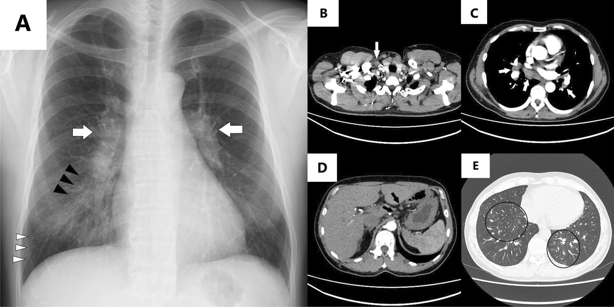
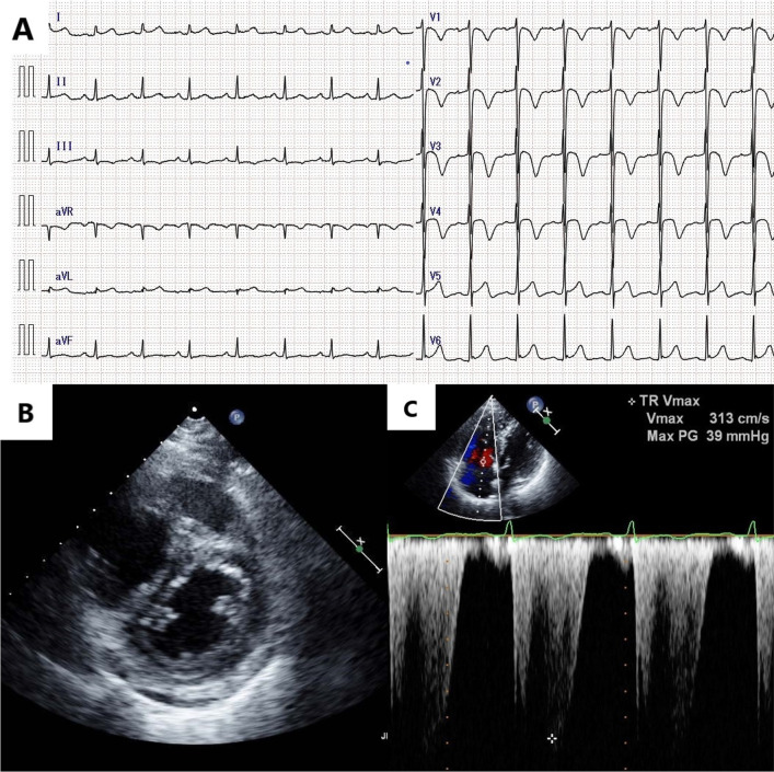
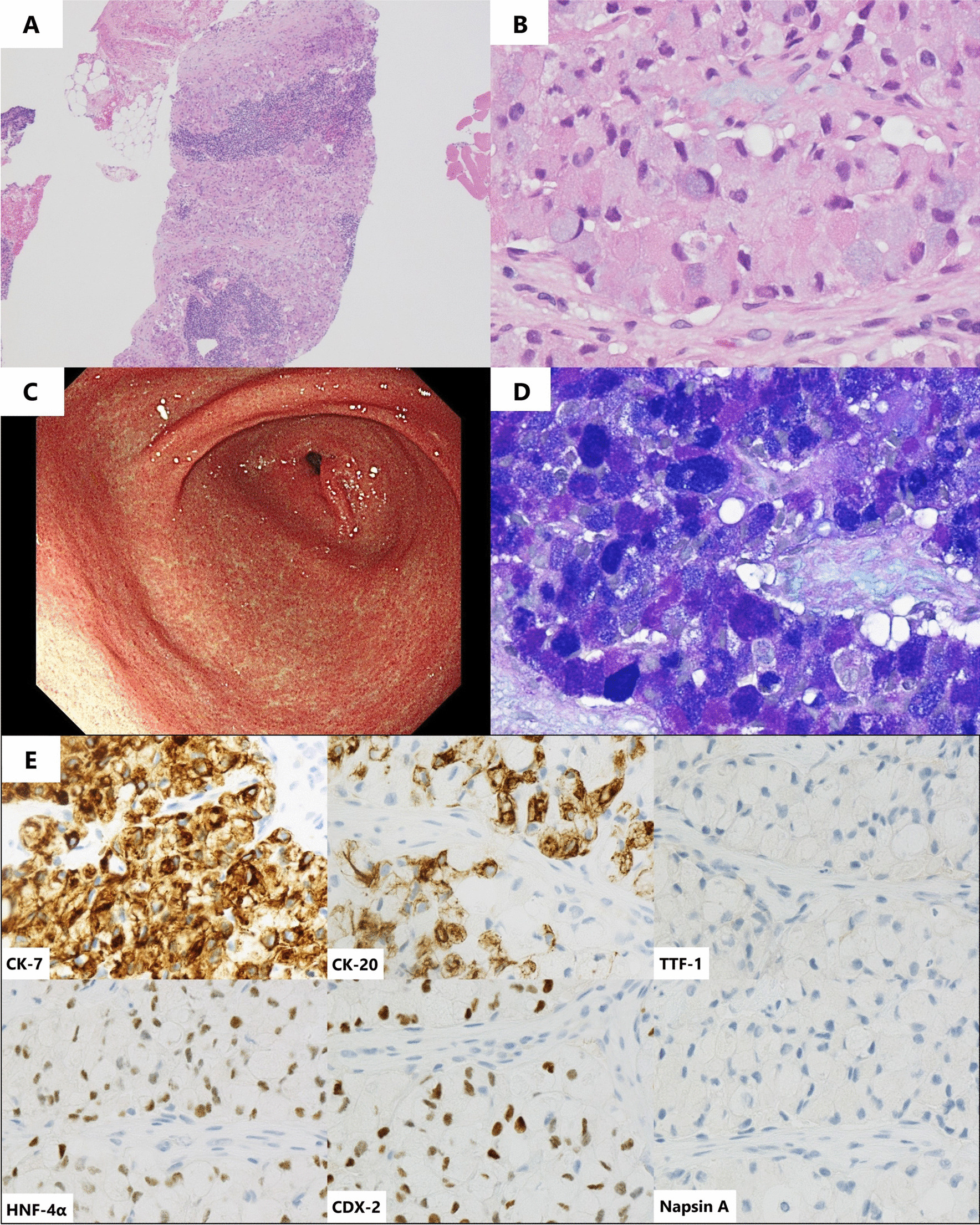
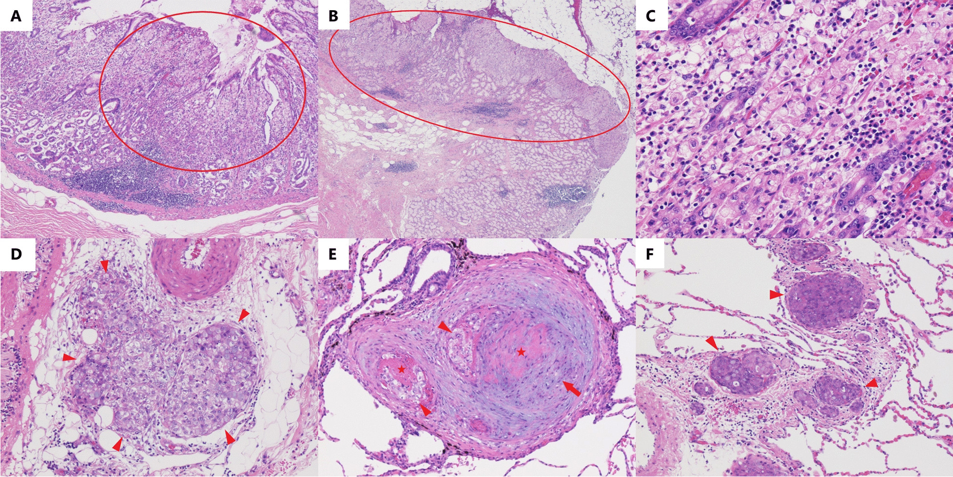
Case presentation: A previously healthy 50-year-old man presented with progressive dyspnea and cough for 1 month. Echocardiography suggested pulmonary hypertension. Computed tomography revealed diffuse lymphadenopathy, whereas blood work revealed an elevation in several serum tumor marker levels. Despite normal upper endoscopic findings, a presumptive diagnosis of PTTM due to gastric cancer was made based on pathological findings of cervical lymph node biopsy, which indicated signet ring cell carcinoma. Imatinib and tegafur/gimeracil/oteracil plus oxaliplatin therapy were started on day 7. The patient's condition was initially stable. However, his symptoms suddenly progressed, and the patient died on day 8. Macroscopic postmortem examination revealed no abnormal gastric wall findings. Microscopically, PTTM was confirmed, and multiple serial sections of the stomach revealed early gastric cancer.

Conclusions: Despite normal endoscopic findings, micro-occult gastric cancer can lead to PTTM. Physicians should be aware of this disease presentation. Taking prompt action is needed when PTTM is suspected, even if the patient appears stable.

Keywords: Early gastric cancer; Endoscopy; Imatinib; Pulmonary tumor thrombotic microangiopathy; Signet ring cell carcinoma.


Conflict of interest statement

The authors declare that there is no competing interests regarding the publication of this article.

Figures

Fig. 1
Fig. 1
Radiographic findings on admission. A Chest radiography shows bilateral hilar lymphadenopathy (white arrows), ground-glass opacities (black arrows), and Kerley B lines (white arrowheads) in the right lung. Enhanced computed tomography shows B right cervical lymphadenopathy (white arrow), C hilar/mediastinal lymphadenopathy (white arrows), and D upper abdominal lymphadenopathy (black arrows). E Septal thickening and ground-glass opacities are observed in the lower lung lobes (black circles)
Fig. 2
Fig. 2
Physiological examination findings on admission. A Electrocardiography shows negative T waves in precordial leads. B Echocardiography shows D-shape of the left ventricle in systolic phase and C elevated tricuspid regurgitation pressure gradient at 39 mmHg
Fig. 3
Fig. 3
Pathological findings of right cervical lymph node biopsy. A Pathological findings of right cervical lymph node biopsy with hematoxylin and eosin staining indicate tumor cells in the specimen (×40) as well as B tumor cells with a signet ring cell feature (×400). C Upper endoscopy shows no abnormal findings in the pyloric region of the stomach. D Lymph node biopsy with Alcian blue-periodic acid-Schiff staining shows mucin-abundant tumor cells (×400). E Immunohistochemical staining shows positive cytokeratin (CK)-7, positive CK-20, negative thyroid transcription factor-1 (TTF-1), positive hepatocyte nuclear factor-4α (HNF-4α), positive caudal-type homeobox-2 (CDX-2), and negative napsin A (×400)
Fig. 4
Fig. 4
Postmortem pathological findings. A, B Postmortem pathological findings of hematoxylin and eosin staining of the stomach show two gastric cancer lesions in the pyloric region (×40 and ×20, respectively). These two lesions are within the lamina propria (circles). C The magnified cancer lesion shows signet ring cell carcinoma (×200). D Lymphovascular invasion is observed in the subserosa (arrowheads) (×100). E Pathological findings of the lung show tumor emboli (arrowheads), thickened epithelial cells (arrow), fibrin thrombi (stars) of small pulmonary arteries (×100), and F carcinomatous lymphangiomatosis (arrowheads) (×100)

References

    1. Price LC, Seckl MJ, Dorfmüller P, Wort SJ. Tumoral pulmonary hypertension. Eur Respir Rev. 2019;28:180065. doi: 10.1183/16000617.0065-2018. - DOI - PMC - PubMed
    1. Godbole RH, Saggar R, Kamangar N. Pulmonary tumor thrombotic microangiopathy: a systematic review. Pulm Circ. 2019;9:2045894019851000. doi: 10.1177/2045894019851000. - DOI - PMC - PubMed
    1. Sato Y, Marutsuka K, Asada Y, Yamada M, Setoguchi T, Sumiyoshi A. Pulmonary tumor thrombotic microangiopathy. Pathol Int. 1995;45:436–440. doi: 10.1111/j.1440-1827.1995.tb03481.x. - DOI - PubMed
    1. Chinen K, Tokuda Y, Fujiwara M, Fujioka Y. Pulmonary tumor thrombotic microangiopathy in patients with gastric carcinoma: an analysis of 6 autopsy cases and review of the literature. Pathol Res Pract. 2010;206:682–689. doi: 10.1016/j.prp.2010.05.002. - DOI - PubMed
    1. Igarashi S, Kawaguchi T, Hoshi N. Early gastric cancer carcinoma with widespread metastasis and pulmonary tumor thrombotic microangiopathy (PTTM): report of an autopsy case. I to Cho (Stomach Intest) 1997;32:861–865.

Publication types

MeSH terms