Review
. 2022 Apr;19(4):228-249.
doi: 10.1038/s41569-021-00629-x. Epub 2021 Nov 10.

Macrophage-targeted nanomedicine for the diagnosis and treatment of atherosclerosis

Affiliations
Review

Macrophage-targeted nanomedicine for the diagnosis and treatment of atherosclerosis

Wei Chen et al. Nat Rev Cardiol. 2022 Apr.

Abstract

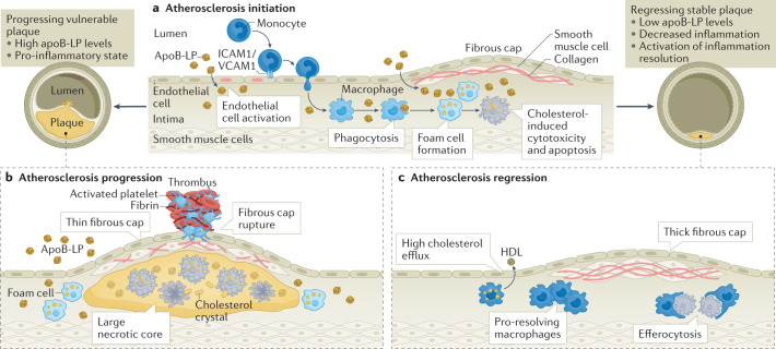
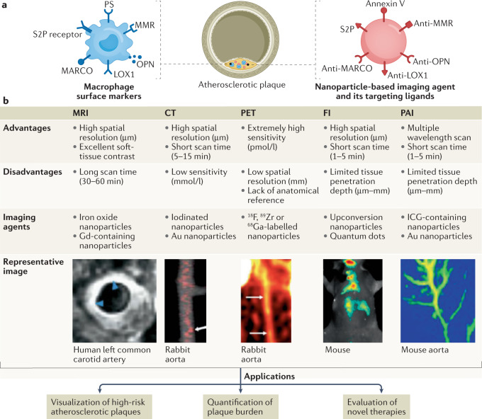
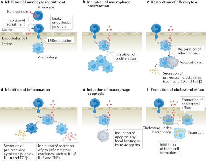
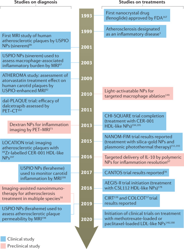
Nanotechnology could improve our understanding of the pathophysiology of atherosclerosis and contribute to the development of novel diagnostic and therapeutic strategies to further reduce the risk of cardiovascular disease. Macrophages have key roles in atherosclerosis progression and, therefore, macrophage-associated pathological processes are important targets for both diagnostic imaging and novel therapies for atherosclerosis. In this Review, we highlight efforts in the past two decades to develop imaging techniques and to therapeutically manipulate macrophages in atherosclerotic plaques with the use of rationally designed nanoparticles. We review the latest progress in nanoparticle-based imaging modalities that can specifically target macrophages. Using novel molecular imaging technology, these modalities enable the identification of advanced atherosclerotic plaques and the assessment of the therapeutic efficacy of medical interventions. Additionally, we provide novel perspectives on how macrophage-targeting nanoparticles can deliver a broad range of therapeutic payloads to atherosclerotic lesions. These nanoparticles can suppress pro-atherogenic macrophage processes, leading to improved resolution of inflammation and stabilization of plaques. Finally, we propose future opportunities for novel diagnostic and therapeutic strategies and provide solutions to challenges in this area for the purpose of accelerating the clinical translation of nanomedicine for the treatment of atherosclerotic vascular disease.


Conflict of interest statement

The authors declare no competing interests.

Figures

Fig. 1
Fig. 1. Role of macrophages in the progression and regression of atherosclerosis.
a | Atherosclerosis development is initiated by the retention and aggregation of apolipoprotein B-containing lipoproteins (apoB-LPs) in the subendothelial space. ApoB-LPs activate endothelial cells, resulting in the upregulation of adhesion molecules (such as intercellular adhesion molecule 1 (ICAM1) and vascular cell adhesion molecule 1 (VCAM1)) that mediate monocyte adhesion to endothelial cells and migration into the arterial vessel wall. In the arterial intima, monocytes differentiate into macrophages, which engulf lipoprotein-derived cholesterol, leading to foam cell formation. Macrophages that cannot process the large amounts of cholesterol undergo cholesterol-induced cytotoxicity and apoptosis. b | If circulating apoB-LP levels remain elevated and the pro-inflammatory state persists, monocyte infiltration and macrophage apoptosis continue, producing so-called ‘vulnerable’ atherosclerotic plaques with large necrotic cores and thin fibrous caps. Vulnerable plaques are prone to rupture, which can lead to thrombus formation, arterial occlusion and sudden cardiac death. c | However, if the plasma cholesterol level is sufficiently lowered and/or the pro-inflammatory state subsides and resolution processes are activated, atherosclerosis regression is possible. Regressing atherosclerotic lesions are characterized by a high cholesterol efflux from macrophages to HDL, large numbers of pro-resolving macrophages that clear apoptotic cells through efferocytosis, reduced plaque necrosis and a thick protective fibrous cap. Regressing atherosclerotic plaques are relatively stable and less likely to rupture than vulnerable plaques and, most importantly, are associated with a lower risk of coronary artery disease in humans.
Fig. 2
Fig. 2. Macrophage-targeted nanoplatforms and non-invasive diagnostic imaging of atherosclerosis.
Non-invasive bioimaging technologies facilitate the visualization of high-risk atherosclerotic plaques with high spatiotemporal resolution. a | The unique epitopes expressed on the macrophage surface can be recognized with the use of targeted nanoparticle-based imaging agents. Targeted nanoparticles have been designed to improve the delivery of imaging agents to inflammatory macrophages in atherosclerotic plaques, thereby improving imaging contrast. b | Various non-invasive bioimaging modalities, advantages, disadvantages and the associated nanoparticle-based imaging contrast agents for plaque visualization. The arrows and arrowheads show areas of high agent uptake. Nanoparticle-facilitated non-invasive bioimaging can provide insights into atherosclerotic plaque biology as well as help to quantify atherosclerosis burden and evaluate the efficacy of therapies at the molecular, cellular and functional levels. FI, fluorescence imaging; ICG, indocyanine green; LOX1, lectin-like oxidized LDL receptor 1; MARCO, macrophage receptor MARCO; MMR, macrophage mannose receptor; OPN, osteopontin; PAI, photoacoustic imaging; PS, phosphatidylserine. MRI image adapted with permission from ref., Elsevier. CT image adapted from ref., Springer Nature Limited. PET image adapted from ref., CC BY 4.0 (https://creativecommons.org/licenses/by/4.0/). FI image adapted with permission from ref., American Chemical Society. PAI image adapted with permission from ref., American Chemical Society.
Fig. 3
Fig. 3. Approaches for atherosclerosis treatment with macrophage-targeting nanotherapeutics.
a | Monocyte recruitment to the atherosclerotic lesion areas can be reduced by delivering therapeutics to monocytes or to vascular endothelial cells by nanoparticles. b | The proliferation of inflammatory macrophages can be inhibited by nanoparticle-assisted delivery of therapeutics to the lesional macrophages. c | The restoration of efferocytosis in macrophages by nanotherapeutics can help to remove dead cells from atherosclerotic plaques, prevent secondary necrosis and elicit the production of anti-inflammatory cytokines such as IL-10 and transforming growth factor-β (TGFβ). d | Inflammation can be ameliorated by modulating the functions of lesional macrophages via nanotherapeutics, increasing the secretion of pro-resolving cytokines (such as IL-10 and TGFβ) or inhibiting the secretion of pro-inflammatory cytokines (such as IL-6, IL-1β and TNF) from lesional macrophages. e | Induction of macrophage apoptosis by local heating or by toxic agents can reduce macrophage burden in atherosclerotic lesions. However, this strategy is suitable only for early lesions. In late atherosclerotic lesions, the impaired phagocytic clearance of apoptotic macrophages might lead to secondary necrosis of these cells and a pro-inflammatory response. f | Promotion of cholesterol efflux from the cholesterol-laden macrophage by nanotherapeutics can reduce foam cell formation.
Fig. 4
Fig. 4. Timeline of major developments in the field of atherosclerosis nanomedicine.
Milestones in the development of nanoparticle-based imaging contrast agents and therapeutics for atherosclerosis diagnosis and treatment. NP, nanoparticle; USPIO, ultrasmall superparamagnetic iron oxide.

References

    1. Virani SS, et al. Heart disease and stroke statistics-2021 update: a report from the American heart association. Circulation. 2021;143:e254–e743. doi: 10.1161/CIR.0000000000000950. - DOI - PubMed
    1. Tabas I, Williams KJ, Borén J. Subendothelial lipoprotein retention as the initiating process in atherosclerosis: update and therapeutic implications. Circulation. 2007;116:1832–1844. doi: 10.1161/CIRCULATIONAHA.106.676890. - DOI - PubMed
    1. Ross R. Atherosclerosis — an inflammatory disease. N. Engl. J. Med. 1999;340:115–126. doi: 10.1056/NEJM199901143400207. - DOI - PubMed
    1. Mestas J, Ley K. Monocyte-endothelial cell interactions in the development of atherosclerosis. Trends Cardiovasc. Med. 2008;18:228–232. doi: 10.1016/j.tcm.2008.11.004. - DOI - PMC - PubMed
    1. Glass CK, Witztum JL. Atherosclerosis. The road ahead. Cell. 2001;104:503–516. doi: 10.1016/S0092-8674(01)00238-0. - DOI - PubMed

MeSH terms