Review
. 2022 Jul;22(7):411-428.
doi: 10.1038/s41577-021-00634-8. Epub 2021 Nov 10.

Crosstalk between the renin-angiotensin, complement and kallikrein-kinin systems in inflammation

Affiliations
Review

Crosstalk between the renin-angiotensin, complement and kallikrein-kinin systems in inflammation

Zivile Bekassy et al. Nat Rev Immunol. 2022 Jul.

Abstract

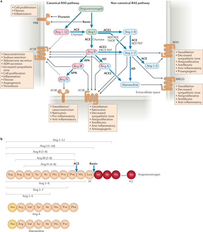
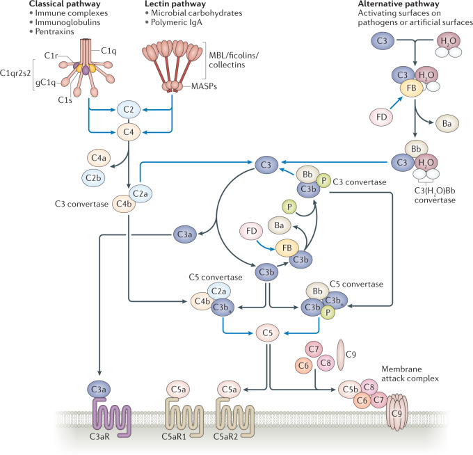
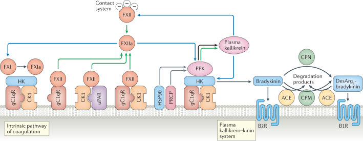
During severe inflammatory and infectious diseases, various mediators modulate the equilibrium of vascular tone, inflammation, coagulation and thrombosis. This Review describes the interactive roles of the renin-angiotensin system, the complement system, and the closely linked kallikrein-kinin and contact systems in cell biological functions such as vascular tone and leakage, inflammation, chemotaxis, thrombosis and cell proliferation. Specific attention is given to the role of these systems in systemic inflammation in the vasculature and tissues during hereditary angioedema, cardiovascular and renal glomerular disease, vasculitides and COVID-19. Moreover, we discuss the therapeutic implications of these complex interactions, given that modulation of one system may affect the other systems, with beneficial or deleterious consequences.


Conflict of interest statement

The authors declare no competing interests.

Figures

Fig. 1
Fig. 1. Overview of the renin–angiotensin system.
a | The renin–angiotensin system (RAS) is initiated by renin, which is secreted by the juxtaglomerular cells of the afferent renal arterioles in response to decreased perfusion, in response to low sodium levels, upon β1-adrenergic receptor stimulation or upon stimulation by nitric oxide (NO) or prostaglandin,. The renin precursor prorenin and renin both bind to prorenin receptor (PRR). In the circulation, renin cleaves angiotensinogen into angiotensin I (Ang I). Prorenin can also cleave angiotensinogen into Ang I, to a certain extent, when bound to PRR. Ang I can also be generated through the cleavage of Ang 1–12 by angiotensin-converting enzyme (ACE) or by chymase in cardiac tissue. The ensuing proteolytic cascade can be divided into the canonical and non-canonical pathways. The canonical pathway is initiated when ACE, or chymase in the heart, cleaves Ang I into Ang II. Binding of Ang II or its cleavage products Ang III or Ang A to the G-protein-coupled receptor Ang II receptor type 1 (AT1R) leads to vasoconstriction, an increase in blood pressure and other effects,. The non-canonical pathway is initiated by Ang I cleavage by ACE2 to Ang 1–9, which is further cleaved into Ang 1–7 (mainly by ACE). In the non-canonical pathway, binding of Ang II, Ang III, Ang 1–9, Ang 1–7 and Ang A to AT2R counteracts effects mediated by AT1R. Ang IV (generated from Ang III when cleaved by aminopeptidase N (APN)) is involved in both the canonical pathway and the non-canonical pathway and binds to angiotensin 4 receptor (AT4R), also known as insulin-regulated aminopeptidase (IRAP). Binding of Ang IV to AT4R has antiapoptotic and both pro-inflammatory and anti-inflammatory effects, as it regulates responses by competitive inhibition of the enzyme/receptor,,. Ang 1–7 binds to the receptors MAS and MAS-related G-protein-coupled receptor (MRGD), and alamandine, which is generated from Ang A or Ang 1–7, also binds to MRGD. Both Ang 1–7 and alamandine have antihypertensive, anti-inflammatory and antifibrotic effects. The non-canonical and canonical RAS pathways can counterbalance each other,–. For example, Ang II binds to both AT2R (non-canonical pathway) and AT1R (canonical pathway) but exerts a more robust effect on AT1R,. AT2R can, however, be upregulated during pathological conditions. Effectors are depicted in circles and the enzymes catalysing their cleavage are listed next to blue arrows. Effectors that predominantly participate in the canonical pathway are coloured pink and those that predominantly participate in the non-canonical pathway are coloured blue. Effectors that participate in both pathways are coloured in both pink and blue, with the predominant colour indicating the more prominent pathway. Black arrows indicate which molecule binds to which receptor, with dashed lines depicting weak receptor binding. b | Amino acid sequences of angiotensin peptides. The cleavage sites of ACE and renin are indicated by blue arrows. AD, aspartate decarboxylase; ADH, antidiuretic hormone; APA, aminopeptidase A; NEP, neprilysin; PEP, prolylendopeptidase; PRCP, prolylcarboxypeptidase; THOP, thimet oligopeptidase.
Fig. 2
Fig. 2. Overview of the complement system.
The complement system is a network that can be divided into the classical, lectin and alternative pathways. The classical pathway is activated when C1s within the C1qr2s2 complex cleaves C2 into C2a and C2b and cleaves C4 into C4a and C4b. Likewise, in the lectin pathway, mannose-binding lectin (MBL) serine protease 1 (MASP1) and MASP2 can also cleave C2 and C4. The classical and lectin pathways converge at the level of C2 and C4 cleavage, where a complex between C2a and C4b forms the C3 convertase. The alternative pathway exhibits a constant low-grade activation on biological surfaces whereby hydrolysed C3, C3(H2O), binds to factor B (FB), which is cleaved by factor D (FD) into the Bb and Ba fragments. The Bb fragment contributes to the formation of the initial convertase C3(H2O)Bb. This convertase cleaves C3 into C3a and C3b. C3b forms a complex with FB, which is again cleaved by FD into Bb, to form C3bBb, the C3 convertase. This convertase is stabilized by properdin (P). The formation of C3 convertases (C4b2a in the classical and lectin pathways and C3bBb in the alternative pathway) is amplified by the C3b-generation amplification loop (centre). By addition of extra C3bs to the C3 convertases, these are transformed into C5 convertases. C5 convertases cleave C5 into C5a and C5b, which promotes the assembly of the pore-forming unit C5b-9 within the cell membrane; this process is called the ‘terminal lytic complement pathway’. Binding of C3a to the receptor C3aR and of C5a to the C5a receptors C5aR1 and C5aR2 induces potent inflammatory responses. Blue lines depict enzymatic activity. Black lines depict binding or conversion into cleavage products. gC1q, globular head of C1q.
Fig. 3
Fig. 3. Overview of the plasma kallikrein–kinin and contact systems.
The plasma kallikrein–kinin and contact systems and their activation on endothelial cells. The zymogen factor XII (FXII) binds to urokinase plasminogen activator receptor (uPAR) in complex with cytokeratin 1 (CK1) or the globular head of the C1q receptor (gC1qR) alone or in complex with CK1 (ref.) and is autoactivated, or is activated by plasma kallikrein, into the enzymatically active FXIIa. Alternatively, in the contact system, FXII is activated to FXIIa when it encounters negatively charged surfaces. Plasma prekallikrein (PPK) is cleaved into plasma kallikrein, its active form, by soluble or bound FXIIa. On the surface of endothelial cells, PPK, when bound to high-molecular-weight kininogen (HK), can also be activated to plasma kallikrein by heat shock protein 90 (HSP90) or by prolylcarboxyeptidase (PRCP) (HSP90 and PRCP act as cofactors rather than enzymes). Thus, prekallikrein can be converted to plasma kallikrein both by FXIIa and by autoactivation in the presence of cofactors. Plasma kallikrein cleaves HK to release the nonapeptide bradykinin, and FXIIa activates FXI by cleaving it to form FXIa, thereby initiating the intrinsic coagulation cascade. Bradykinin exerts inflammatory effects via the B2 receptor (B2R). Bradykinin is degraded to desArg9-bradykinin by carboxypeptidase M (CPM) on the cell surface or by carboxypeptidase N (CPN) in the fluid phase. DesArg9-bradykinin binds preferentially to the B1 receptor (B1R). Both kinins are further degraded by angiotensin-converting enzyme (ACE). Green arrows show that autoactivation of a protein into its activated form can occur, blue arrows indicate enzymatic activity, black arrows point to cleavage products and grey arrows indicate cofactors.
Fig. 4
Fig. 4. Interactions between the renin–angiotensin, complement and kallikrein–kinin systems on the vascular wall.
a | Joint pathways of the renin–angiotensin system (RAS), complement system and kallikrein–kinin system (KKS) on the endothelium. Certain components of the complement system and the KKS bind to the same surface receptor, the globular head of C1q receptor (gC1qR). It can bind C1q or, in complex with cytokeratin 1 (CK1), it binds high-molecular-weight kininogen (HK) and plasma prekallikrein (PPK). C1 inhibitor is a common inhibitor of plasma kallikrein, activated factor XII (FXIIa), mannose-binding lectin (MBL) serine proteases (MASPs), C1s and C1r. Plasma kallikrein cleaves HK, releasing bradykinin. In vitro it was also shown to cleave complement factor B (FB) and C3; however, this has not been confirmed in vivo. Renin cleaves both angiotensinogen into angiotensin I (Ang I) and C3 into C3a and C3b allowing formation of more C3bBb. Angiotensin-converting enzyme (ACE) degrades bradykinin to metabolites and also converts Ang I to Ang II. Red lines depict inhibition, green arrows indicate that autoactivation of a protein can occur, blue arrows indicate enzymatic activity (dashed if shown only in vitro) and black arrows point to cleavage products. b | Mechanisms of vascular permeability and leakage induced by the RAS, the KKS and the complement system. Vascular permeability via the RAS is induced by Ang II signalling via Ang II type 1 receptor (AT1R). In the KKS, vasopermeability is associated with bradykinin and desArg9-bradykinin signalling via the receptors B2R and B1R, respectively, and in the complement system with C5a signalling via the C5a receptor (C5aR1) and deposition of C5b-9 within the cell membrane. c | Mechanisms of endothelial cell injury, thrombosis, vasopermeability/oedema and inflammation induced by the RAS, the KKS/contact system and the complement system during severe acute respiratory syndrome coronavirus 2 (SARS-CoV-2) infection. After binding to pulmonary epithelial cells via surface-expressed ACE2, SARS-CoV-2 undergoes endocytosis, thereby reducing the amount of ACE2 on cell surfaces. This creates an imbalance in the RAS with increased activation of the canonical pathway. ACE2 inactivates desArg9-bradykinin. Decreased levels of ACE2 therefore increase desArg9-bradykinin levels and signalling via B1R in the KKS, leading to inflammation. SARS-CoV-2 interacts with C1 inhibitor, the level of which was found to be reduced in bronchoalveolar lavage fluid from patients with COVID-19 (ref.). Downregulation of C1 inhibitor will allow activation of the classical and lectin pathways of the complement system, and the nucleocapsid proteins of SARS-CoV and SARS-CoV-2 were shown to bind to MASP2 and initiate the lectin pathway of the complement system. The KKS/contact system is also activated because C1 inhibitor inhibits activated factor XII (FXIIa). Furthermore, activated FXII cleaves C1r, activating the classical pathway of the complement system. Endothelial cells become injured through excess activation of the three pathways. Endothelial cell activation leads to increased vascular permeability (as shown in part b), resulting in pulmonary oedema. Ang II, C3a and C5a as well as the kinin effectors bradykinin and desArg9-bradykinin promote neutrophil recruitment and inflammation. Neutrophil proteases can further activate the KKS by cleavage of HK. As listed in Table 1, effectors of the KKS and FXII, effectors of the RAS canonical pathway and of the complement system induce thrombosis. uPAR, urokinase plasminogen activator receptor.

References

    1. Santos RAS, et al. The renin-angiotensin system: going beyond the classical paradigms. Am. J. Physiol. Heart Circ. Physiol. 2019;316:H958–H970. - PMC - PubMed
    1. Noris M, Remuzzi G. Overview of complement activation and regulation. Semin. Nephrol. 2013;33:479–492. - PMC - PubMed
    1. Colman RW, Schmaier AH. Contact system: a vascular biology modulator with anticoagulant, profibrinolytic, antiadhesive, and proinflammatory attributes. Blood. 1997;90:3819–3843. - PubMed
    1. Forrester SJ, et al. Angiotensin II signal transduction: an update on mechanisms of physiology and pathophysiology. Physiol. Rev. 2018;98:1627–1738. - PMC - PubMed
    1. Li F, Li W, Farzan M, Harrison SC. Structure of SARS coronavirus spike receptor-binding domain complexed with receptor. Science. 2005;309:1864–1868. - PubMed

Publication types

Substances