

Proliferation and Differentiation of Dopaminergic Neurons from Human Neuroepithelial Stem Cells Obtained from Embryo Reduction Following In Vitro Fertilization
- PMID: 34759448
- PMCID: PMC8563046
- DOI: 10.5455/medarh.2021.75.280-285
Proliferation and Differentiation of Dopaminergic Neurons from Human Neuroepithelial Stem Cells Obtained from Embryo Reduction Following In Vitro Fertilization
Abstract
Background: Recent advances in stem cell technologies have rekindled an interest in the use of cell therapies to treat patients with Parkinson's disease. Although the transplantation of dopaminergic mesencephalic human fetal brain tissue has previously been reported in the treatment of patients with Parkinson's disease, this method is limited by the availability of tissue obtained from each human embryo.
Objective: Our study aimed to isolate, culture, proliferate, and differentiate dopaminergic neurons from human neuroepithelial stem cells obtained from embryo reduction procedures performed in multifetal pregnancies following in vitro fertilization.
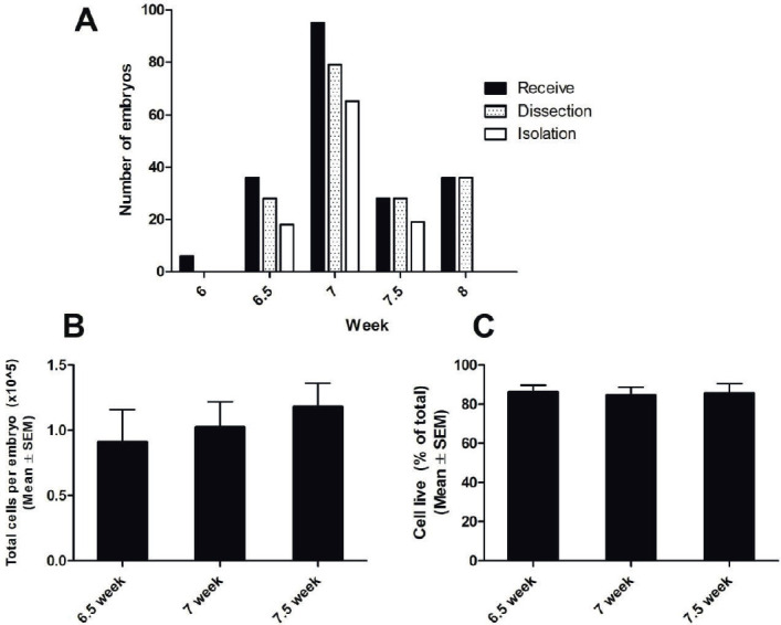
Materials and methods: A total of 201 human embryos were dissected for isolation and culture of neuroepithelial stem cells for proliferation and differentiation into dopaminergic neurons. All embryos were obtained from embryo reduction procedures performed in multifetal pregnancies after in vitro fertilization treatments.
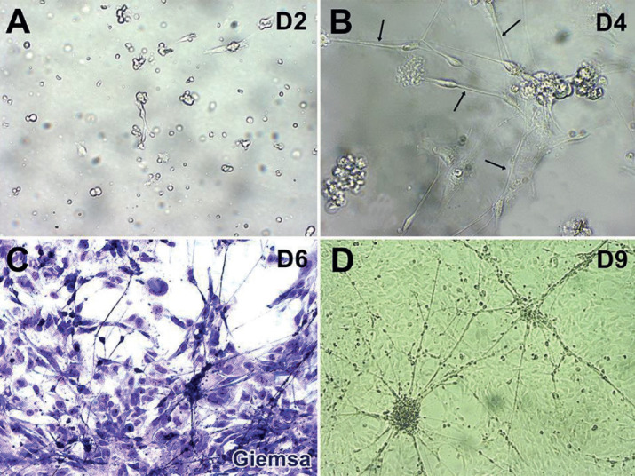
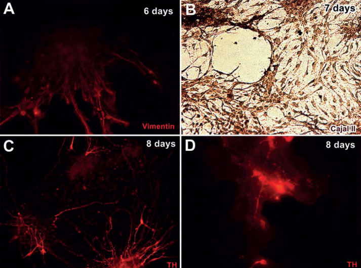
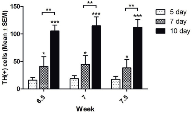
Results: Human neuroepithelial stem cells were isolated and cultured from embryos from 6.0 to 8.0 weeks. Neuroepithelial stem cells were successfully isolated, proliferated, and differentiated into dopaminergic neurons. The cells adhered to the surfaces of cell culture plates after 2 days and could be proliferated and differentiated into neurons within 4 days. Cultured cells expressed the dopaminergic marker tyrosine hydroxylase after 6 days, suggesting that these cells were successfully differentiated into dopaminergic neurons.
Conclusion: The successful isolation, culture, proliferation, and differentiation of human dopaminergic neurons from embryo reductions performed for multifetal pregnancies after in vitro fertilization suggests that this pathway may serve as a potential source of cell therapy materials for use in the treatment of Parkinson's disease.
Keywords: Parkinson’s disease; dopaminergic neuron; human neuroepithelial stem cells; in vitro fertilization.
© 2021 Nguyen Thi Binh, Nguyen Khang Son, Dao Thuy Phuong, Do Thuy Huong, Nguyen Phuc Hoan, Nguyen Thanh Hoa, Nguyen Minh Duc, Nguyen Manh Ha.
Conflict of interest statement
There are no conflicts of interest to declare.
Figures
References
-
- Pires AO, Teixeira FG, Mendes-Pinheiro B, Serra SC, Sousa N, Salgado AJ. Old and new challenges in Parkinson’s disease therapeutics. Prog Neurobiol. 2017;156:69–89. - PubMed
-
- Lindvall O. Clinical translation of stem cell transplantation in Parkinson’s disease. J Intern Med. 2016;279(1):30–40. - PubMed
-
- Perlow MJ, Freed WJ, Hoffer BJ, Seiger A, Olson L, Wyatt RJ. Brain grafts reduce motor abnormalities produced by destruction of nigrostriatal dopamine system. Science. 1979;204(4393):643–647. - PubMed
-
- Barker RA, Barrett J, Mason SL, Bjorklund A. Fetal dopaminergic transplantation trials and the future of neural grafting in Parkinson’s disease. Lancet Neurol. 2013;12(1):84–91. - PubMed
-
- Freed CR, Greene PE, Breeze RE, et al. Transplantation of embryonic dopamine neurons for severe Parkinson’s disease. N Engl J Med. 2001;344(10):710–719. - PubMed
Publication types
MeSH terms
LinkOut - more resources
Full Text Sources