

Late diagnosis of hepatic mesenchymal hamartoma and placental mesenchymal dysplasia
- PMID: 34760455
- PMCID: PMC8409553
- DOI: 10.1002/ajum.12021
Late diagnosis of hepatic mesenchymal hamartoma and placental mesenchymal dysplasia
Abstract
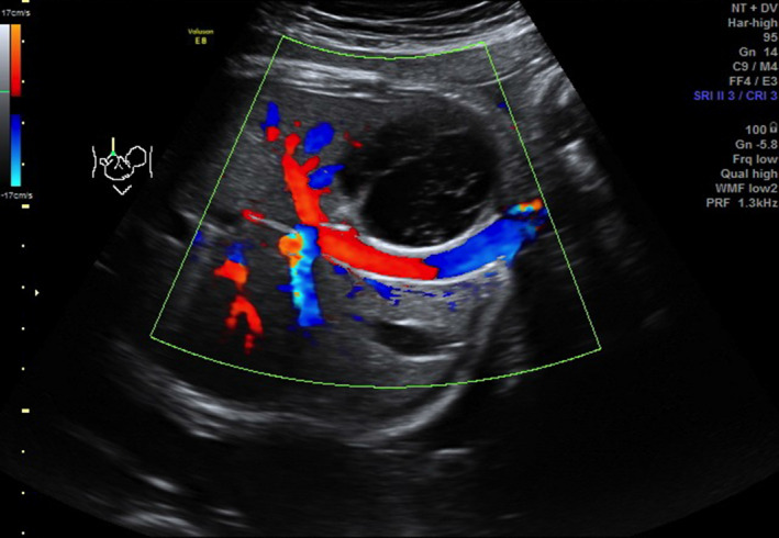
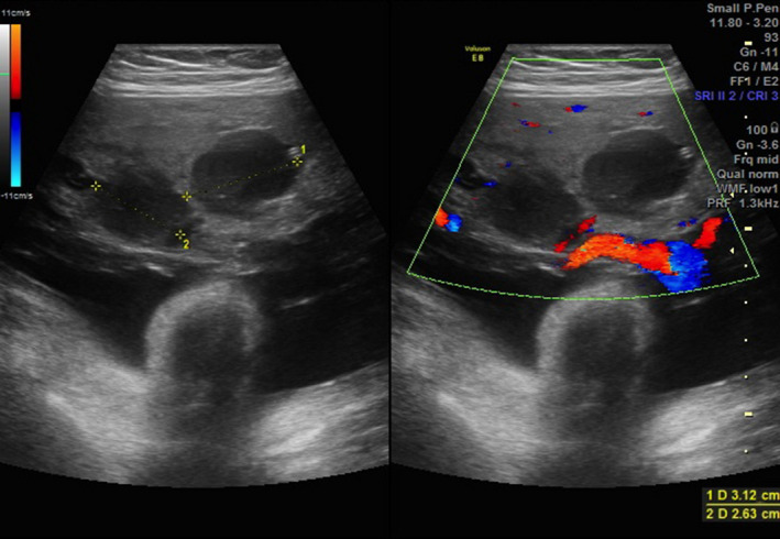
Placental mesenchymal dysplasia (PMD) is a rare condition characterised by placental enlargement, oedematous villi and multiple anechoic cysts. Hepatic mesenchymal hamartoma (HMH) is a benign proliferation of mesenchymal tissue, commonly seen in infants below the age of 2. We report the case of a 28 years old female who was noted to have a fetus with a well-circumscribed cyst on the liver, suggestive of HMH and a large, thickened placenta, with multiple anechoic cysts, consistent with PMD during the third trimester. There were no other structural abnormalities and at 38 weeks she underwent an induction of labour with normal vaginal delivery of a live female infant. While the aetiology is poorly understood, the increased incidence of HMH with PMD and the morphological similarities of the changes seen in both the placenta and liver, suggests a possible common developmental mechanism. There are only 12 other cases of this concurrent pathology in the literature and only one of these had resulted in a term delivery, and ours is the second one to date.
Keywords: anechoic cysts; fetal liver cysts; hepatic mesenchymal hamartoma; placental mesenchymal dysplasia.
© 2016 Australasian Society for Ultrasound in Medicine.
Figures


References
-
- Kinoshita T, Fukaya S, Yasuda Y, Itoh M. Placental mesenchymal dysplasia. J Obstet Gynaecol Res 2007; 33(1): 83–6. - PubMed
-
- Lennington WJ, Gray GF Jr, Page DL. Mesenchymal hamartoma of liver. A regional ischemic lesion of a sequestered lobe. Am J Dis Child 1993; 147(2): 193–6. - PubMed
-
- Nayeri U, West A, Grossetta Nardini H, Copel JA, Sfakianaki AK. Systematic review of sonographic findings of placental mesenchymal dysplasia and subsequent pregnancy outcomes. Ultrasound Obstet Gynecol 2013; 41(4): 366–74. - PubMed
Publication types
LinkOut - more resources
Full Text Sources