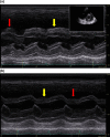
Paradoxical septal motion: A diagnostic approach and clinical relevance
- PMID: 34760507
- PMCID: PMC8409894
- DOI: 10.1002/ajum.12086
Paradoxical septal motion: A diagnostic approach and clinical relevance
Abstract
Abnormal septal motion (commonly referred to as septal bounce) is a common echocardiographic finding that occurs with several conditions, including the following: mitral stenosis, left bundle branch block, pericardial syndromes and severe pulmonary hypertension. We explore the subtle changes that occur on M-mode imaging of the septum, other associated echocardiographic features, the impact of inspiratory effort on septal motion and relevant clinical findings. Finally, we discuss the impact of abnormal septal motion on cardiac form and function, proposing there is a clinically significant impact on biventricular filling and ejection.
Keywords: cardiac pacing; cardiac surgery; cardiology.
© 2018 Australasian Society for Ultrasound in Medicine.
Figures


References
-
- Kaul S. The interventricular septum in health and disease. Am Heart J 1986; 112: 568–81. - PubMed
-
- Grines CL, Bashore TM, Boudoulas H, Olson S, Shafer P, Wooley CF. Functional abnormalities in isolated left bundle branch block. The effect of interventricular asynchrony. Circulation 1989; 79(4): 845–53. - PubMed
-
- Dillon JC, Chang S, Feigenbaum H. Echocardiographic manifestations of left bundle branch block. Circulation 1974; 49: 876–80. - PubMed
-
- Little WC, Reeves RC, Arciniegas J, Katholi RE, Rogers EW. Mechanism of abnormal interventricular septal motion during delayed left ventricular activation. Circulation 1982; 65(7): 1486–91. - PubMed
-
- Walmsley J, Huntjens PR, Prinzen FW, Delhaas T, Lumens J. Septal flash and septal rebound stretch have different underlying mechanisms. Am J Physiol Heart Circ Physiol 2016; 310(3): H394–403. - PubMed
Publication types
LinkOut - more resources
Full Text Sources
