. 2021 Oct 25:11:735277.
doi: 10.3389/fonc.2021.735277. eCollection 2021.

A Role of CXCL1 Drives Osteosarcoma Lung Metastasis via VCAM-1 Production

Affiliations

A Role of CXCL1 Drives Osteosarcoma Lung Metastasis via VCAM-1 Production

Chiang-Wen Lee et al. Front Oncol. .

Abstract

Osteosarcoma, a common aggressive and malignant cancer, appears in the musculoskeletal system among young adults. The major cause of mortality in osteosarcoma was the recurrence of lung metastases. However, the molecular mechanisms of metastasis involved in osteosarcomas remain unclear. Recently, CXCL1 and CXCR2 have been crucial indicators for lung metastasis in osteosarcoma by paracrine releases, suggesting the involvement of directing neutrophils into tumor microenvironment. In this study, overexpression of CXCL1 has a positive correlation with the migratory and invasive activities in osteosarcoma cell lines. Furthermore, the signaling pathway, CXCR2/FAK/PI3K/Akt, is activated through CXCL1 by promoting vascular cell adhesion molecule 1 (VCAM-1) via upregulation of nuclear factor-kappa B (NF-κB) expression and nuclear translocation. The in vivo animal model further demonstrated that CXCL1 serves as a critical promoter in osteosarcoma metastasis to the lung. The correlated expression of CXCL1 and VCAM-1 was observed in the immunohistochemistry staining from human osteosarcoma specimens. Our findings demonstrate the cascade mechanism regulating the network in lung metastasis osteosarcoma, therefore indicating that the CXCL1/CXCR2 pathway is a worthwhile candidate to further develop treatment schemas.

Keywords: CXCL1; VCAM-1; metastasis; migration; osteosarcoma.


Conflict of interest statement

The authors declare that the research was conducted in the absence of any commercial or financial relationships that could be construed as a potential conflict of interest.

Figures
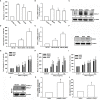
Figure 1
Figure 1
CXCL1 increased migration, invasion, and wound healing in human osteosarcoma MG63, U2OS, and HOS cells. (A) The cell migration ability of the osteoblast cell line hFOB 1.19 and the osteosarcoma cell lines MG63, U2OS, and HOS was assessed using the Transwell assay. (B, C) Total mRNA and protein were collected from the indicated cell lines, and CXCL1 expression was detected using qPCR and Western blotting. (D–F) Migratory ability and CXCL1 expression of the indicated cells (MG63, M10, and M20) were examined by Transwell, qPCR, and immunoblotting assays. (G–I) Cells were incubated with CXCL1 (1–10 ng/ml) and the Transwell assay were used for detecting in vitro migratory, wound healing, and invasive activities after 18 h. (J–L) Cells were transfected with pcDNA3.1-conjugated CXCL1 plasmid and then CXCL1 mRNA, protein expression, and migratory potential were measured by immunoblotting, qPCR, and Transwell assays. Results were expressed as mean ± S.D. from at least three individual experiments. *p < 0.05 compared with the control group of each experiment.
Figure 2
Figure 2
CXCL1 promotes osteosarcoma migration by increasing VCAM-1 expression. (A) MG63 cells were stimulated with CXCL1 (1–10 ng/ml) and the mRNA levels of VCAM-1 and ICAM-1 were examined by real-time qPCR. (B, C) CXCL1 increased the VCAM-1 mRNA and protein expression in MG63, U2OS, and HOS human osteosarcoma cells. (D) VCAM-1 expression and migratory potential were examined by immunoblotting and Transwell assays in MG63 cells, which were transfected with VCAM-1 shRNA for 24 h and then stimulated with CXCL1 for another 24 h. The data were collected from at least three individual experiments and expressed as mean ± S.D. *p < 0.05 as compared to the control group; # p < 0.05 compared with the CXCL1-treated shRNA-control group.
Figure 3
Figure 3
The role of the CXCR2 receptor in CXCL1-mediated cell migration and VCAM-1 expression. (A, B) The CXCR2 antagonist, SB225002. (C, D) CXCR2-shRNA transfection. (E, F) CXCR2 neutralizing antibody reduced CXCL1-induced cell migration and VCAM-1 expression in MG63 cells. The data were collected from at least three individual experiments and expressed as mean ± S.D. *p < 0.05 as compared to the control group; # p < 0.05 compared with the CXCL1-treated shRNA-control and IgG groups.
Figure 4
Figure 4
The FAK, PI3K, Akt, and NF-κB pathways are involved in CXCL1-promoted migration and VCAM-1 expression in MG63 osteosarcoma cell. (A, B) CXCL1-increased cell migration and VCAM-1 mRNA expression could be reduced by pre-treated FAK inhibitor (2 μM), PI3K inhibitors LY294002 (5 μM) and wortmannin (2 μM), Akt inhibitor (5 μM), and NF-κB inhibitors TPCK (5 μM) and PDTC (1 μM) for 90 min. (C) Phosphorylated levels of FAK, P85, and Akt, and (D) phosphorylated levels of IKK, IκBα, and p65 were examined by immunoblotting assay in MG-63 cells with CXCL1 incubation for the indicated time intervals (0, 10, 15, 30, or 60 min). (E, F) Transfecting dominant-negative mutants of FAK, PI3K, Akt, and IKK for 24 h could suppress CXCL1 increased cell migration and VCAM-1 mRNA expression. (G–I) FAK, PI3K, Akt, and NF-κB inhibitors reduced CXCL1-induced VCAM-1 protein expressions. The data were collected from at least three individual experiments and expressed as mean ± S.D. *p < 0.05 as compared to the control group; # p < 0.05 compared with the CXCL1-treated control or vector group.
Figure 5
Figure 5
NF-κB mediates the response of human osteosarcoma cells by CXCL1 stimulation. (A) The luciferase activity of NF-κB promoter plasmid was incubated with the indicated doses of CXCL1 for 24 h in MG63 cells. CXCL1 (1-50 ng/ml)-stimulated NF-κB luciferase activity was suppressed by (B) inhibitors of FAK, PI3K, and AKT and (C) dominant negative FAK, PI3K, AKT, IKK, or CXCR2 shRNA. (D) The anti-p65 and DAPI signals by pretreated several inhibitors and then CXCL1 (10 ng/ml)-stimulated, representative confocal microscopy images were shown. (E) CXCL1-induced p65 phosphorylation and (F) chromatin immunoprecipitation of anti-p65 in nuclear extracts of MG63 cells were measured with pretreated inhibitors. One percent of immunoprecipitated chromatin was assayed to verify equal loading (input). Results are expressed as the mean ± S.D. of triplicate samples. *p < 0.05 as compared to the control group; # p < 0.05 compared with the CXCL1-treated control or vector group.
Figure 6
Figure 6
The CXCL1/CXCR2 axis is required for osteosarcoma lung metastasis. (A, B) Protein and mRNA levels of CXCL1 and (C) cell migration ability of MG63 transfected with sh-CXCLl by immunoblotting, real-time qPCR, and Transwell assays in MG63 cells. (D) The mice were injected with MG63/control shRNA, or MG63/CXCL1 shRNA cells. Lung metastasis was monitored by bioluminescence imaging at the indicated time intervals. (E–G) The appearance of hematoxylin and eosin stain and numbers of metastasis nodules of lung specimens from sacrifice mice after 4 weeks of cell injection was shown. Results are expressed as the mean ± S.D. of triplicate samples. */# p < 0.05 as compared to the shRNA-control group..
Figure 7
Figure 7
Clinical significance of CXCL1 and VCAM-1 in osteosarcoma patient specimens. (A) CXCL1 and VCAM-1 expression levels in specimens were determined in a tumor tissue microarray by IHC staining. The stained specimens were photographed using an optical microscope. (B, C) IHC stain intensities were scored from 1 to 5 to quantify CXCL1 and VCAM-1 expression levels in different stages. (D) Correlation between CXCL1 and VCAM-1 expression in human osteosarcoma specimens. Results are shown as mean ± S.D.

References

    1. Ottaviani G, Jaffe N. The Etiology of Osteosarcoma. Cancer Treat Res (2009) 152:15–32. doi: 10.1007/978-1-4419-0284-9_2 - DOI - PubMed
    1. Arndt CA, Crist WM. Common Musculoskeletal Tumors of Childhood and Adolescence. N Engl J Med (1999) 341(5):342–52. doi: 10.1056/NEJM199907293410507 - DOI - PubMed
    1. Fan TM, Roberts RD, Lizardo MM. Understanding and Modeling Metastasis Biology to Improve Therapeutic Strategies for Combating Osteosarcoma Progression. Front Oncol (2020) 10:13. doi: 10.3389/fonc.2020.00013 - DOI - PMC - PubMed
    1. Isakoff MS, Bielack SS, Meltzer P, Gorlick R. Osteosarcoma: Current Treatment and a Collaborative Pathway to Success. J Clin Oncol (2015) 33(27):3029–35. doi: 10.1200/JCO.2014.59.4895 - DOI - PMC - PubMed
    1. Ando K, Mori K, Verrecchia F, Marc B, Redini F, Heymann D. Molecular Alterations Associated With Osteosarcoma Development. Sarcoma (2012) 2012:523432. doi: 10.1155/2012/523432 - DOI - PMC - PubMed