Review
. 2021 Nov 8:9:tkab035.
doi: 10.1093/burnst/tkab035. eCollection 2021.

Deciphering glial scar after spinal cord injury

Affiliations
Review

Deciphering glial scar after spinal cord injury

Yu Zhang et al. Burns Trauma. .

Abstract

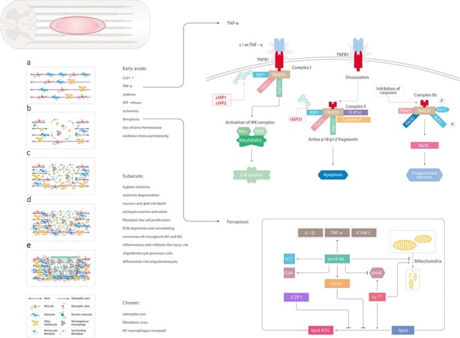
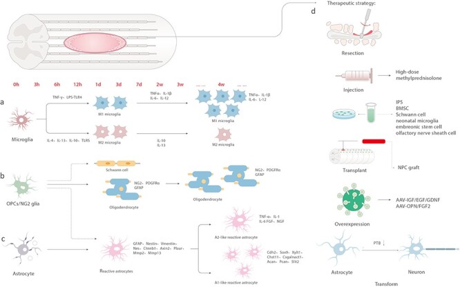
Spinal cord injury (SCI) often leads to permanent disability, which is mainly caused by the loss of functional recovery. In this review, we aimed to investigate why the healing process is interrupted. One of the reasons for this interruption is the formation of a glial scar around the severely damaged tissue, which is usually covered by reactive glia, macrophages and fibroblasts. Aiming to clarify this issue, we summarize the latest research findings pertaining to scar formation, tissue repair, and the divergent roles of blood-derived monocytes/macrophages, ependymal cells, fibroblasts, microglia, oligodendrocyte progenitor cells (OPCs), neuron-glial antigen 2 (NG2) and astrocytes during the process of scar formation, and further analyse the contribution of these cells to scar formation. In addition, we recapitulate the development of therapeutic treatments targeting glial scar components. Altogether, we aim to present a comprehensive decoding of the glial scar and explore potential therapeutic strategies for improving functional recovery after SCI.

Keywords: Axon regeneration; Glial scar; Spinal cord injury; Therapeutic strategy.


Figures

Figure 1.
Figure 1.
Schematic illustration of the main changes induced by spinal cord injury. (a) Uninjured spinal cord. (b) Early acute phase of spinal cord injury: increased level of Ca2+, TNF-α, edema, ATP release, ischemia, ferroptosis, loss of ionic homeostasis, oxidative stress excitotoxicity. Soluble TNF-α binds to TNFR1 on the cell membrane. RIP1 binds to other related proteins to form complex 1. cIAP1 and cIAP2 can ubiquitinate RIP1 and activate the NF-κB signaling pathway to promote cell survival. USP21 induces deubiquitination of RIP1 to form complex 2, leading to apoptosis. Fe2+ catalyzes the peroxidation of liposomes on the cell membrane and increases the production of reactive oxygen species. In addition, GPX4 is inactivated, thereby inducing iron-dependent death of cells. The iron-dependent death inhibitor, SRS 16–86, significantly inhibits the expression of inflammatory factors IL-1β, TNF-α and ICAM-1 and rescues the reduction of mitochondria and crest reduction. (c, d) Subacute phase: hypoxic ischemia, reactive astrocytosis, Wallarian degeneration, neurons and glial cell death, reactive activation of astrocytes, fibroblast-like cell proliferation, ECM deposition and remodeling, conversion of microglia to M1 and M2, infiltration of inflammatory cells at the injury site, differentiation of OPCs into oligodendrocytes. (e) Chronic phase: formation of astrocyte scars, fibroblast scars and increase in M1 macrophages. TNF- α tumor necrosis factor-alpha, ATP adenosine triphosphate, RIP1 receptor interacting protein 1, cIAP1 apoptosis inhibitory protein 1, GPX4 glutathione peroxidase 4, IL-1β interleukin-1β, ICAM-1 intercellular adhesion molecule-1, OPCs oligodendrocyte progenitor cells
Figure 2.
Figure 2.
Temporal changes and treatment in spinal cord injury. (a) IFN-γ and LPS-TLR4 induce microglia to switch to M1 type within 3 days of injury and secrete TNF-α, IL-1β, IL-6 and IL-12. IL-4, IL-13, IL-10 and TLRs stimulate the switch to M2 type, which secrete IL-10 and IL-13. The M1 type eventually dominates. (b) OPCs/NG2 can differentiate into Schwann cells and astrocytes in the early stage of injury, or these can differentiate into oligodendrocytes and express NG2, PDGFRα and GFAP. (c) Astrocytes transform into reactive astrocytes after injury and up-regulate the expression of GFAP, nestin, vimentin, Nes, Ctnnb1, Axin2, Plaur, Mmp2 and Mmp13; however, over 2–4 weeks, A1-like astrocytes appear and up-regulate Cdh2, Sox9, Xylt1, Chst11, Csgalnact1, Acan, Pcan, Slit2 and another type of scar that secretes TNF-α, IL-1, IL-6, FGF and NGF. (d) Therapeutic strategy: surgical treatment, high-dose methylprednisolone, cell (iPS, BMSC, Schwann cell, neonatal microglia, embryonic stem cells, olfactory nerve sheath cells) transplantation, cocktail therapy, transforming astrocytes into neurons. IFN-γ interferon-gamma, TNF- α tumor necrosis factor-alpha, IL interleukin, OPCs oligodendrocyte progenitor cells, GFAP glial fibrillary acidic protein, FGF fibroblast growth factor, NGF nerve growth factor

References

    1. Courtine G, Sofroniew MV. Spinal cord repair: advances in biology and technology. Nat Med. 2019;25:898–908. - PubMed
    1. He Y, Liu X, Chen Z. Glial scar-a promising target for improving outcomes after CNS injury. J Mol Neurosci. 2020;70:340–52. - PubMed
    1. Yu B, Yao C, Wang Y, Mao S, Wang Y, Wu R, et al. The landscape of gene expression and molecular regulation following spinal cord Hemisection in rats. Front Mol Neurosci. 2019;12:287. - PMC - PubMed
    1. Adams KL, Gallo V. The diversity and disparity of the glial scar. Nat Neurosci. 2018;21:9–15. - PMC - PubMed
    1. Sami A, Selzer ME, Li S. Advances in the Signaling pathways downstream of glial-scar axon growth inhibitors. Front Cell Neurosci. 2020;14:174. - PMC - PubMed