. 2021 Nov 3;16(1):1642-1652.
doi: 10.1515/med-2021-0357. eCollection 2021.

miR-150-5p affects AS plaque with ASMC proliferation and migration by STAT1

Affiliations

miR-150-5p affects AS plaque with ASMC proliferation and migration by STAT1

Yuan Bian et al. Open Med (Wars). .

Abstract

We explore miR-150-5p in atherosclerosis (AS). The AS model was constructed using Apo E-/- mice with an injection of the miR-150-5p mimic or an inhibitor. Pathological characteristics were assessed using Oil red O staining and Masson staining. Quantitative reverse transcription-polymerase chain reaction (qRT-PCR) and Western blot were used to analyze the expressions of microRNA-150-5p (miR-150-5p), STAT1, α-SMA (α-smooth muscle actin) and proliferating cell nuclear antigen (PCNA). Targetscan and dual-luciferase reporter assay were used to analyze the interaction between miR-150-5p and STAT1. The viability, migration, cell cycle and α-SMA and PCNA expressions in oxidized low-density lipoprotein (ox-LDL)-stimulated primary human aortic smooth muscle cells (ASMCs) were assessed using molecular experiments. miR-150-5p was reduced in both AS mice and ox-LDL-stimulated human aortic smooth muscle cells but STAT1 had the opposite effect. The miR-150-5p inhibitor alleviated the increase of lipid plaque and reduced collagen accumulation in the aortas during AS. Upregulation of α-SMA and PCNA was reversed by miR-150-5p upregulation. STAT1 was targeted by miR-150-5p, and overexpressed miR-150-5p weakened the ox-LDL-induced increase of viability and migration abilities and blocked cell cycle in ASMCs, but overexpressed STAT1 blocked the effect of the miR-150-5p mimic. This paper demonstrates that miR-150-5p has potential as a therapeutic target in AS, with plaque stabilization by regulating ASMC proliferation and migration via STAT1.

Keywords: atherosclerosis; collagen metabolism; miR-150-5p; plaque stability; signal transducer and activator of transcription 1.


Conflict of interest statement

Conflict of interest: The authors declare no conflicts of interest.

Figures

Figure 1
Figure 1
miR-150-5p regulated plaque stability, collagen metabolism and expressions of proliferation biomarkers in ApoE−/− mice. (a) Transfection efficiency of the miR-150-5p mimic and inhibitor in the aortas of ApoE−/− mice was detected using qPCR. U6 snRNA was used as the internal reference for normalization of miR-150-5p levels. (b) The level of lipid plaques in aortas of ApoE−/− mice was determined by Oil red O staining. Magnification: 100×, scale bar = 100 μm. (c) Masson staining was performed to assess the collagen accumulation in aortas of ApoE−/− mice. Magnification: 200×, scale bar = 50 μm. (d)–(h) Western blot analysis of α-SMA, PCNA and STAT1 expressions in aorta tissues. GAPDH was used as the internal reference. Three mice were in each group. *** p < 0.001 vs control; # p < 0.05, ### p < 0.001 vs Model + NC. NC: negative control for the mimic and inhibitor. M: miR-150-5p mimic. I: miR-150-5p inhibitor. All these experiments were performed three times independently. qRT-PCR: quantitative reverse transcription-polymerase chain reaction; ox-LDL: oxidized low-density lipoprotein; M: miR-150-5p mimic; NC: negative control (empty vector).
Figure 2
Figure 2
STAT1 was a direct target gene of miR-150-5p. (a) Targetscan predicted that miR-150-5p might bind to STAT1 mRNA. (b) The interaction between miR-150-5p and STAT1 was confirmed by using a dual-luciferase reporter assay. HASMCs were co-transfected with the recombinant plasmid containing the STAT1 WT sequence (WT) or mutant sequence (MUT) with or without the miR-150-5p mimic. M: miR-150-5p mimic; Blank: cells were treated with PBS without transfection. *** p < 0.001 vs Blank. The luciferase reporter assay was repeated three times.
Figure 3
Figure 3
miR-150-5p regulated the expression of STAT1 in ox-LDL-stimulated HASMCs. (a) The relative expression of miR-150-5p was detected by qRT-PCR. U6 snRNA was used as the internal reference for normalization of miR-150-5p levels. (b)–(d) The relative expression of STAT1 was detected by qRT-PCR and Western blot. GAPDH was used as the internal reference. ** p < 0.01,*** p < 0.001 vs Control; ### p < 0.001 vs ox-LDL + NC; ^^ p < 0.01, ^^^ p < 0.001 vs ox-LDL + M; §§§ p < 0.001 vs ox-LDL + STAT1. qRT-PCR: quantitative reverse transcription-polymerase chain reaction; ox-LDL: oxidized low-density lipoprotein; M: miR-150-5p mimic; NC: negative control (empty vector). All these experiments were performed three times independently.
Figure 4
Figure 4
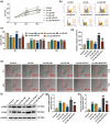
miR-150-5p influenced the viability, migration and expressions of proliferation biomarkers of ox-LDL-stimulated HASMCs via STAT1. (a) CCK-8 assay was used to determine the viability of ox-LDL-stimulated HASMCs transfected with the mimic or overexpressed STAT1 at 0 h, 24 h and 48 h. (b and c) Flow cytometry was used to detect the cell cycle of ox-LDL-stimulated HASMCs transfected with mimic or overexpressed STAT1. (d) The relative migration rate normalized to the control group was calculated. (e) Wound healing assay was performed to assess the cell migration ability. Magnification: 100×, scale bar = 50 μm. (f)–(h) Western blot analysis of α-SMA and PCNA in ox-LDL-stimulated HASMCs transfected with the mimic or overexpressed STAT1. GAPDH was used as the internal reference. ** p < 0.01,*** p < 0.001 vs Control; # p < 0.05, ## p < 0.01, ### p < 0.001 vs ox-LDL + NC; ^ p < 0.05, ^^ p < 0.01, ^^^ p < 0.001 vs ox-LDL + mimic; § p < 0.05, §§ p < 0.01, §§§ p < 0.001 vs ox-LDL + STAT1. All these experiments were performed three times independently. OD: optical density; M: miR-150-5p mimic; NC: negative control (empty vector); ox-LDL: oxidized low-density lipoprotein; HASMCs: human aortic smooth muscle cells; α-SMA, α-smooth muscle actin (α-SMA); PCNA: proliferating cell nuclear antigen; STAT1: transducer and activator of transcription STAT 1.

References

    1. Lüscher TF, Landmesser U, von Eckardstein A, Fogelman AM. High-density lipoprotein: vascular protective effects, dysfunction, and potential as therapeutic target. Circ Res. 2014;114(1):171–82. - PubMed
    2. Lüscher TF, Landmesser U, von Eckardstein A, Fogelman AM. High-density lipoprotein: vascular protective effects, dysfunction, and potential as therapeutic target. Circ Res. 2014;114(1):171–82. - PubMed
    1. Glass CK, Witztum JL. Atherosclerosis. the road ahead. Cell. 2001;104(4):503–16. - PubMed
    2. Glass CK, Witztum JL. Atherosclerosis. the road ahead. Cell. 2001;104(4):503–16. - PubMed
    1. Billon C, Sitaula S, Burris TP. Inhibition of RORα/γ suppresses atherosclerosis via inhibition of both cholesterol absorption and inflammation. Mol Metab. 2016;5(10):997–1005. - PMC - PubMed
    2. Billon C, Sitaula S, Burris TP. Inhibition of RORα/γ suppresses atherosclerosis via inhibition of both cholesterol absorption and inflammation. Mol Metab. 2016;5(10):997–1005. - PMC - PubMed
    1. Falk E. Pathogenesis of atherosclerosis. J Am Coll Cardiol. 2006;47(8 Suppl):C7–12. - PubMed
    2. Falk E. Pathogenesis of atherosclerosis. J Am Coll Cardiol. 2006;47(8 Suppl):C7–12. - PubMed
    1. Bennett MR, Sinha S, Owens GK. Vascular smooth muscle cells in atherosclerosis. Circ Res. 2016;118(4):692–702. - PMC - PubMed
    2. Bennett MR, Sinha S, Owens GK. Vascular smooth muscle cells in atherosclerosis. Circ Res. 2016;118(4):692–702. - PMC - PubMed

LinkOut - more resources