. 2021 Oct 14;24(11):103275.
doi: 10.1016/j.isci.2021.103275. eCollection 2021 Nov 19.

An exercise "sweet spot" reverses cognitive deficits of aging by growth-hormone-induced neurogenesis

Affiliations

An exercise "sweet spot" reverses cognitive deficits of aging by growth-hormone-induced neurogenesis

Daniel G Blackmore et al. iScience. .

Abstract

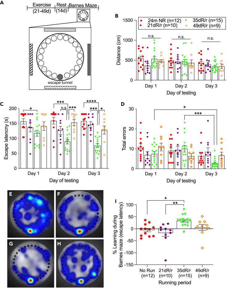
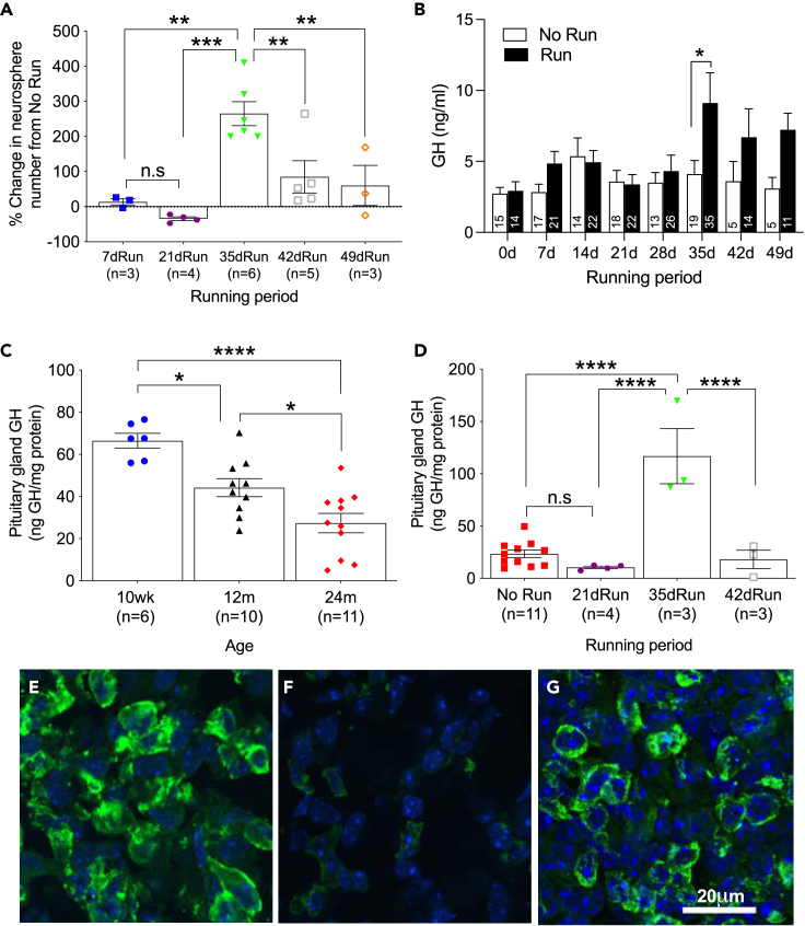
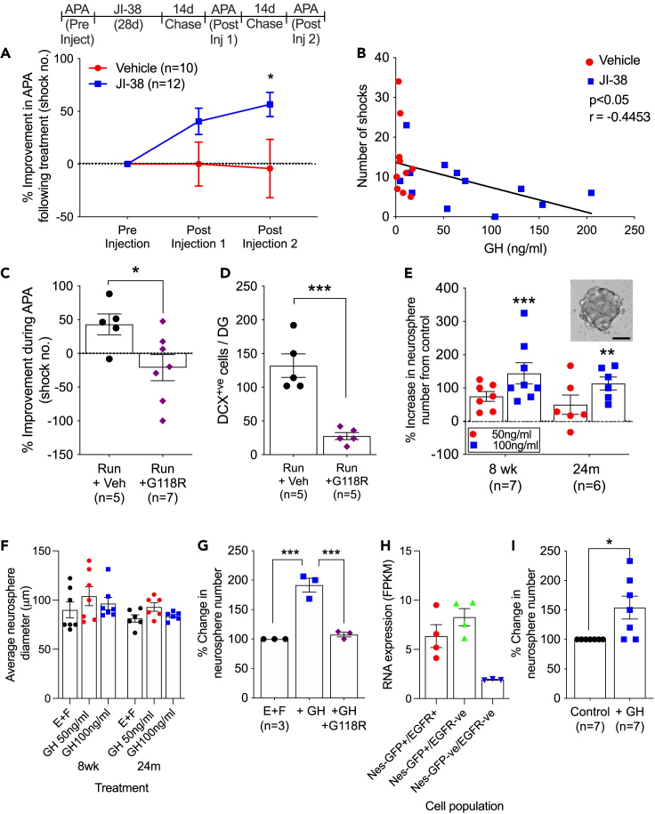
Hippocampal function is critical for spatial and contextual learning, and its decline with age contributes to cognitive impairment. Exercise can improve hippocampal function, however, the amount of exercise and mechanisms mediating improvement remain largely unknown. Here, we show exercise reverses learning deficits in aged (24 months) female mice but only when it occurs for a specific duration, with longer or shorter periods proving ineffective. A spike in the levels of growth hormone (GH) and a corresponding increase in neurogenesis during this sweet spot mediate this effect because blocking GH receptor with a competitive antagonist or depleting newborn neurons abrogates the exercise-induced cognitive improvement. Moreover, raising GH levels with GH-releasing hormone agonist improved cognition in nonrunners. We show that GH stimulates neural precursors directly, indicating the link between raised GH and neurogenesis is the basis for the substantially improved learning in aged animals.

Keywords: Age; Endocrine system physiology; Neuroscience.


Conflict of interest statement

The authors declare no competing interests.

Figures

None
Graphical abstract
Figure 1
Figure 1
Aged animals display a spatial learning impairment that is ameliorated by an optimized period of physical exercise (A) Representation of the APA apparatus. Each mouse is placed on a rotating grid and must use the spatial cues located around the room to avoid the stationary shock zone (shown in red). (B) Naïve mice from 10 weeks to 24 months of age were tested on the APA task. The 18- and 24-month-old animals were unable to learn as evidenced by the lack of change in shock number over the test period (mean ± SE; two-way RM-ANOVA [age effect F(4, 119) = 50.79; p < 0.0001] with Bonferroni post hoc tests). (C) During APA testing, there was a significant difference in the total distance traveled for the different age groups; however, on the final day, there were no significant differences between the groups (mean ± SE; two-way RM-ANOVA age effect [F(4,119) = 1.750; p = 0.1435], with Bonferroni post hoc tests. ∗ represents significance between 10 weeks and 18 months old; # represents significance between 12 months old and 18 months old). (D) Schematic representation of the experimental design. All 24-month-old animals were tested for spatial learning ability prior to exercise. At the completion of the exercise period, the running wheels were removed. Two weeks later, novel cues were used in the second test of spatial learning. (E) The running time for group-housed animals for 21 d, 35 d, or 49 d revealed no difference in average daily running time during the exercise intervention (mean ± SE). Analysis of running activity was calculated between the hours of 7 pm and 3 am, when the majority of exercise took place. (F) Only the 24-month-old animals that underwent the optimized exercise period of 35 days improved during APA testing, to a similar level to that of young animals (mean ± SE; two-way RM-ANOVA [exercise effect F(7, 100) = 9.208; p < 0.0001] with Bonferroni post hoc tests). (G) There was a significant increase in maximal time spent avoiding the shock zone for the 35-d run group, and on the final day, this was significantly different from the time recorded for all other groups (mean ± SE; two-way RM-ANOVA [exercise effect [F(6,89) = 4.723, p = 0.0003], with Bonferroni post hoc tests). (H) The learning ability following exercise was calculated by comparing the number of shocks on the first day of testing to the number of shocks on the last day of testing, which was represented as a percentage (mean ± SE; one-way ANOVA [F(6,89) = 3.092; p = 0.0085], with Bonferroni post hoc tests). This revealed that the animals that ran for 35 days significantly improved whereas those that underwent other exercise periods did not. (I) There was no difference and no improvement in the number of shocks received during APA testing for animals that had access to a stationary running wheel (mean ± SE; two-way RM-ANOVA, with Bonferroni post hoc tests). ∗p < 0.05, ∗∗p < 0.01, ∗∗∗∗p < 0.0001. NR, no run, R/r, Run/rest where rest is 14 days. See also Figure S1.
Figure 2
Figure 2
The optimal period of exercise improves Barnes maze performance in 24-month-old animals (A) Schematic of the experimental approach to test animals following exercise with the Barnes maze. Each animal used spatial cues placed evenly around the room to find and enter the escape tunnel (marked in gray). Three trials per day for 3 days were conducted, with each animal placed in a different quadrant to start each trial. (B) There was no difference between groups for the total distance traveled during testing. (C) The 35-d runners were the only group to exhibit a significant reduction in the time to enter the escape tunnel during the course of testing (mean ± SE; two-way RM-ANOVA [exercise effect F(3,42) = 13.32 p < 0.0001], with Bonferroni post hoc tests). (D) Only the 35-d runner group exhibited significantly fewer errors during the testing period (mean ± SE; two-way RM-ANOVA [F(6,84) = 2.77; p = 0.0163], with Bonferroni post hoc tests). (E–H) Heat maps reveal differences in the areas of the Barnes maze explored prior to escape on the final day of testing for (E) no run, (F) 21-d run, (G) 35-d run, and (H) 49-d run animals, with hotter colors representing more time spent in that position. (I) There was a significant increase in learning ability for animals that ran for 35 d (mean ± SE; one-way ANOVA [F(3,42) = 6.109; p = 0.0015], with Bonferroni post hoc tests). ∗p < 0.05, ∗∗p < 0.01, ∗∗∗p < 0.001, ∗∗∗∗p < 0.0001. NR, no run, R/r, Run/rest where rest is 14 days.
Figure 3
Figure 3
Exercise increases the frequency of immature neurons required for spatial learning in the aged brain (A) The total number of DCX+ve cells in the DG decreases with age (mean ± SE; one-way ANOVA [F (4,39) = 80.55, p < 0.0001] with Bonferroni post hoc tests). (B) Following cognitive testing, only those 24-month-old animals that underwent exercise for 35 d showed a significant increase in DCX+ve cells in the DG relative to controls and all other running periods (mean ± SE; one-way ANOVA [F(6,77) = 8.255; p < 0.0001] with Bonferroni post hoc tests). (C–E) Representative photomicrographs of the DG of (C) 24-m no-run, (D) 24-m 21-d run, or (E) 24-m 35-d run mice, labeled for DCX (green) and DAPI (blue). Scale bar = 50 μm. Arrowheads point to DCX+ve cells. (F) 24m 35dR/14dr animals show an increase in branching morphology compared with age-matched no run controls (mean ± SE; two-way ANOVA [F(6,64) = 4.939; p < 0.0003] with Bonferroni post hoc tests). (G) Schematic representation of the experimental design to specifically ablate DCX+ve cells following exercise. At the end of the running period, animals were injected with either vehicle or DT and then tested on the APA task. The number of shocks received by DCXDTR animals injected with DT did not decrease during APA testing, whereas the number received by mice injected with vehicle did (mean ± SE; two-way RM-ANOVA [effect of treatment; F(2,49) = 3.767; p = 0.0301], with Bonferroni post hoc tests). There was a significant difference in the shock number on the final day of testing between Run DCXDTR injected with DT and DCXDTR run animals injected with vehicle and WT run animals injected with DT. (H and I) (H) 24-month-old DCXDTR animals injected with DT failed to learn and had significantly fewer DCX+ve cells in the DG compared with control animals (I) (mean ± SE; one-way ANOVA [F(2,49) = 6.381, p = 0.0034] and [F(2,33) = 8.101; p = 0.0014], respectively, with Bonferroni post hoc tests). ∗p < 0.05, ∗∗p < 0.01, ∗∗∗p < 0.001, ∗∗∗∗p < 0.0001. NR, no run; R/r Run/rest where rest is 14 days. See also Figures S2 and S3.
Figure 4
Figure 4
Changes in growth hormone levels during exercise mirror the timing for the activation of endogenous hippocampal precursor cells (A) 35 d of exercise significantly increased hippocampal neurosphere numbers relative to age-matched no-run controls and for all other running periods (no-run controls standardized to zero, mean ± SE; one-way ANOVA [F (5,21) = 13.32; p < 0.0001] with Bonferroni post hoc test). (B) There was a significant increase in GH in the blood of 24-month-old animals following 35 d of exercise (mean + SE; multiple t tests; n values are given in each column of the graph). (C) Naïve pituitary gland GH levels decreased during aging (mean ± SE; one-way ANOVA [F (2,24) = 16.57; p < 0.0001] with Bonferroni post hoc tests). (D) Pituitary gland GH levels showed a significant increase following 35 d of exercise in 24-month-old mice (mean ± SE; one-way ANOVA [F (3,17) = 22.99, p < 0.0001] with Bonferroni post hoc tests). (EG) Representative photomicrographs of pituitary gland cells labeled for GH (green) and DAPI (blue) in (E) 10-wk naive, (F) 24-m naive and (G) 24-m 35-d run mice (Scale bar = 20 μm). ∗p < 0.05, ∗∗p < 0.01; ∗∗∗p < 0.001; ∗∗∗∗p < 0.0001. See also Figures S4–S6.
Figure 5
Figure 5
Pharmacological induction of GH release with JI-38 results in improved cognition in aged animals (A) Comparison of the learning ability prior to treatment and following JI-38 administration revealed a continued improvement in spatial learning, reaching significance at the second postinjection test period (mean ± SE; two-way RM-ANOVA [F(2,40) = 3.243; p = 0.0495] with Bonferroni post hoc tests). (B) Following JI-38 treatment, there was a significant correlation, with higher circulating GH levels correlating to a lower number of shocks received on the final day of APA testing. (C) Animals infused with the GH antagonist G118R during exercise showed no improvement in APA testing (mean ± SE; Student’s t test; t score = 2.421, p = 0.036). (D) Infusion of G118R during exercise prevented an increase in DCX+ve cell number in the DG (mean ± SE; Student’s t test; t score = 5.749, p = 0.0004). (E) The addition of 100 ng/mL of GH significantly increased primary hippocampal neurosphere numbers for both young and 24-month-old animals (mean ± SE; one-way ANOVA [F(2,19) = 11.17; p = 0.0006 and [F(2, 15) = 7.907; p = 0.0045], relative to control (E + F) conditions respectively, with Bonferroni post hoc tests). Insert shows a representative neurosphere. Scale bar = 50 μm. (F) There was no change in neurosphere diameter with the addition of GH. (G) The GH antagonist G118R prevented the GH-dependent increase in hippocampal neurospheres (mean ± SE; one-way ANOVA [F(2,6) = 50.87; p = 0.0002] with Bonferroni post hoc tests). (H) GHR RNA was expressed on the purified, precursor cell population (NesGFP+ve/EGFR+ve). (I) Addition of 100 ng/mL of GH to the purified, clonal hippocampal precursor cells significantly increased neurosphere number (mean ± SE; Student’s t test; t score = 2.782, p = 0.0166). ∗p < 0.05, ∗∗p < 0.01, ∗∗∗p < 0.001. See also Figure S8.
Figure 6
Figure 6
Manipulation of somatostatin alters growth hormone levels and behavior in 24-month-old mice (A) Circulating somatostatin levels decreased after 35 and 42 d of exercise but returned to control levels after 49 d of running (mean ± SE; one-way ANOVA [F(5,56) = 2.436; p = 0.045], with Bonferroni post hoc tests). (B) Somatostatin levels were reduced 2 weeks after the optimized exercise period relative to no run controls and animals that ran beyond 35 d (mean ± SE; one-way ANOVA [F(2,15) = 4.5; p = 0.0295], with Bonferroni post hoc tests). (C) GH levels were elevated at 49 d with continued running when animals were injected daily for 14 d with donepezil (mean + SE; one-way ANOVA [F(2,35) = 4.858; p = 0.0137], with Bonferroni post hoc tests) compared with noninjected 49-d runners (49-d run GH data from Figure 4). (D) Analysis of performance during APA testing revealed a significant improvement only for those animals injected with donepezil during exercise (mean ± SE; one-way ANOVA [F2,35) = 4.8; p = 0.014], with Bonferroni post hoc tests). (E) There was a significant increase in DCX+ve cell number in animals that received donepezil during the final 14 days of exercise (mean ± SE; one-way ANOVA [F2,26) = 8.239; p = 0.0017], with Bonferroni post hoc tests). p < 0.05 Run/r: Run/rest where r refers to a rest period of 14 days. See also Figure S9.

References

    1. Aberg D.N., Lind J., Isgaard J., Georg Kuhn H. Peripheral growth hormone induces cell proliferation in the intact adult rat brain. Growth Horm. IGF Res. 2010;20:264–269. - PubMed
    1. Ajo R., Cacicedo L., Navarro C., Sanchez-Franco F. Growth hormone action on proliferation and differentiation of cerebral cortical cells from fetal rat. Endocrinology. 2003;144:1086–1097. - PubMed
    1. Alba M., Schally A.V., Salvatori R. Partial reversibility of growth hormone (GH) deficiency in the GH-releasing hormone (GHRH) knockout mouse by postnatal treatment with a GHRH analog. Endocrinology. 2005;146:1506–1513. - PubMed
    1. Altman J., Das G.D. Autoradiographic and histological evidence of postnatal hippocampal neurogenesis in rats. J. Comp. Neurol. 1965;124:319–335. - PubMed
    1. Ben Abdallah N.M., Slomianka L., Vyssotski A.L., Lipp H.P. Early age-related changes in adult hippocampal neurogenesis in C57 mice. Neurobiol. Aging. 2010;31:151–161. - PubMed

LinkOut - more resources