. 2022 Feb;31(2):371-383.
doi: 10.1002/pro.4229. Epub 2021 Nov 20.

Three-dimensional structure of xylonolactonase from Caulobacter crescentus: A mononuclear iron enzyme of the 6-bladed β-propeller hydrolase family

Affiliations

Three-dimensional structure of xylonolactonase from Caulobacter crescentus: A mononuclear iron enzyme of the 6-bladed β-propeller hydrolase family

Johan Pääkkönen et al. Protein Sci. 2022 Feb.

Abstract

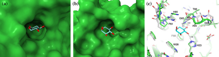
Xylonolactonase Cc XylC from Caulobacter crescentus catalyzes the hydrolysis of the intramolecular ester bond of d-xylonolactone. We have determined crystal structures of Cc XylC in complex with d-xylonolactone isomer analogues d-xylopyranose and (r)-(+)-4-hydroxy-2-pyrrolidinone at high resolution. Cc XylC has a 6-bladed β-propeller architecture, which contains a central open channel having the active site at one end. According to our previous native mass spectrometry studies, Cc XylC is able to specifically bind Fe2+ . The crystal structures, presented here, revealed an active site bound metal ion with an octahedral binding geometry. The side chains of three amino acid residues, Glu18, Asn146, and Asp196, which participate in binding of metal ion are located in the same plane. The solved complex structures allowed suggesting a reaction mechanism for intramolecular ester bond hydrolysis in which the major contribution for catalysis arises from the carbonyl oxygen coordination of the xylonolactone substrate to the Fe2+ . The structure of Cc XylC was compared with eight other ester hydrolases of the β-propeller hydrolase family. The previously published crystal structures of other β-propeller hydrolases contain either Ca2+ , Mg2+ , or Zn2+ and show clear similarities in ligand and metal ion binding geometries to that of Cc XylC. It would be interesting to reinvestigate the metal binding specificity of these enzymes and clarify whether they are also able to use Fe2+ as a catalytic metal. This could further expand our understanding of utilization of Fe2+ not only in oxidative enzymes but also in hydrolases.

Keywords: Caulobacter crescentus; crystal structure; enzyme mechanism; hydrolase; iron; metal coordination; metalloenzyme; xylonolactonase; β-propeller hydrolase.


Conflict of interest statement

The authors declare that they have no competing interests.

Figures

FIGURE 1
FIGURE 1
(a) The hydrolysis of d‐xylonolactone, which is suggested to involve the isomerization reaction of d‐xylono‐1,4‐lactone to d‐xylono‐1,5‐lactone as the first step, which is catalyzed non‐enzymatically by a divalent metal ion (M2+). The lactonase‐catalyzed hydrolysis reaction produces d‐xylonic acid, which has relatively low pK a (see also Figure 5). (b) The ligands successfully used as cryoprotectants and observed in the active site of the enzyme
FIGURE 2
FIGURE 2
(a) The overall three‐dimensional structure of the Cc XylC. The polypeptide chain of monomeric protein is presented as a green cartoon model. The blades of the β‐propeller are numbered from 1 to 6. The bound Fe2+ ion in the central channel is shown as a brown ball and d‐xylopyranose as a stick model. (b) The superimposed eight monomers from the three crystal structures are shown as grey ribbon chains. The additional bound ligand molecules are shown as green stick models
FIGURE 3
FIGURE 3
The active site and bound ligands. The protein structures are shown as green stick models and ligand structures as cyan stick models. Iron atoms and water oxygens are presented as balls in brown and red, respectively. The lengths of hydrogen bonds and coordination bonds are given in ångströms. The side chains from three amino acid residues (Glu18, Asn146, and Asp196), which are bound to the Fe2+, are all in the same plane (planar triad). (a) The bound d‐xylopyranose in the structure of the rectangular crystal, molecule A. (b) The bound HPD in the crystal structure, molecule B. (c) The active site without the ligand in the crystal structure with HPD, molecule A. (d) Modelled binding of d‐xylono‐1,5‐lactone (in yellow) to the active site (same as in c). (e) Modelled binding of d‐xylono‐1,4‐lactone (in yellow) to the active site (same as in c). (f) All three experimental structures superimposed together with the models of two isomers of d‐xylonolactone (in yellow)
FIGURE 4
FIGURE 4
The binding of d‐xylopyranose anomers to the Cc XylC. Ligands are shown as cyan stick models and protein surfaces in green. (a) β‐d‐Xylopyranose binding to the active site. (b) α‐d‐Xylopyranose binding to the putative allosteric site on the protein surface. (c) Details of the putative allosteric site. The protein atoms in complex with α‐d‐xylopyranose are in green, and without the ligand in grey
FIGURE 5
FIGURE 5
The proposed reaction mechanism for the Cc XylC catalyzed hydrolysis of d‐xylono‐1,5‐lactone to form d‐xylonic acid. See the text for more details
FIGURE 6
FIGURE 6
Superimposition of representative members (Table 2) of the 6‐bladed β‐propeller hydrolase family. (a) Cartoon representation of protein monomers. The Cc XylC is in green, other proteins are in grey. The active site ligands are shown as stick models. (b) The superimposition of metal coordination in active sites. The proteins that have three amino acids in plane, which participate in metal ion binding, are in green (xylonolactonase, mouse and human SMP30, and luciferin‐regenerating enzyme), and proteins that have four amino acids in plane are in cyan (Drp35, gluconolactonase, diisopropylfluorophosphatase, and paraoxonase). The interacting residues in the Cc XylC are labelled as E18, D196, and N146. (c,d) The detailed comparison of coordination of catalytic metal ion in Cc XylC (c), and mouse SMP30 (d). The bond lengths between the metal ion and ligand atoms are given in ångströms. Carbon atoms of proteins are in green and of bound ligand (xylose in C, and 1,5‐anhydro‐d‐glucitol in d) in cyan. Both crystal structures have been determined at 1.7 Å resolution. The comparison shows a clear resemblance in metal binding

References

    1. Boer H, Andberg M, Pylkkänen R, Maaheimo H, Koivula A. In vitro reconstitution and characterisation of the oxidative d‐xylose pathway for production of organic acids and alcohols. AMB Expr. 2019;9:48. - PMC - PubMed
    1. Buchert J, Viikari L. The role of xylonolactone in xylonic acid production by Pseudomonas fragi . Appl Microbiol Biotechnol. 1988;27:333–336.
    1. Toivari M, Nygård Y, Kumpula E‐P, et al. Metabolic engineering of Saccharomyces cerevisiae for bioconversion of d‐xylose to d‐xylonate. Metab Eng. 2012;14:427–436. - PubMed
    1. Stephens C, Christen B, Fuchs T, Sundaram V, Watanabe K, Jenal U. Genetic analysis of a novel pathway for d‐xylose metabolism in Caulobacter crescentus . J Bacteriol. 2007;189:2181–2185. - PMC - PubMed
    1. Jermyn MA. Studies on the glucono‐δ‐lactonase of Pseudomonas fluorescens . Biochim Biophys Acta. 1960;37:78–92. - PubMed

Publication types