

An improved procedure for isolating adult mouse cardiomyocytes for epicardial activation mapping
- PMID: 34761519
- PMCID: PMC8650026
- DOI: 10.1111/jcmm.17049
An improved procedure for isolating adult mouse cardiomyocytes for epicardial activation mapping
Abstract
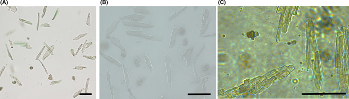
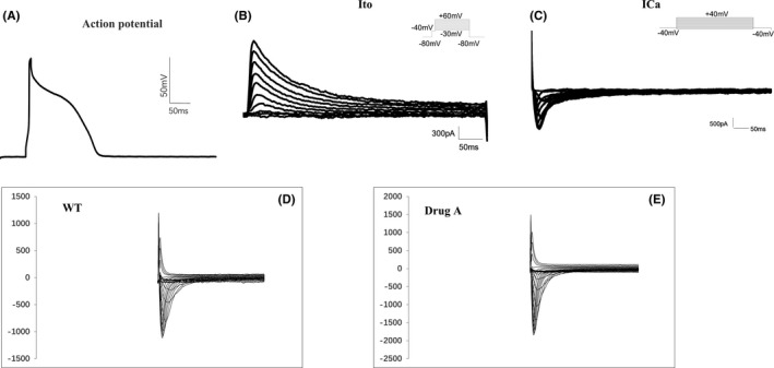
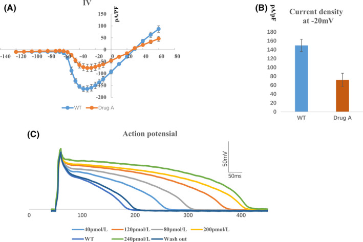
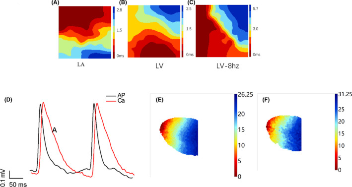
Cardiovascular disease is a leading cause of death and disability worldwide. Although genetically modified mouse models offer great potential for robust research in vivo, in vitro studies using isolated cardiomyocytes also provide an important approach for investigating the mechanisms underlying cardiovascular disease pathogenesis and drug actions. Currently, isolation of mouse adult cardiomyocytes often relies on aortic retrograde intubation under a stereoscopic microscope, which poses considerable technical barriers and requires extensive training. Although a simplified, Langendorff-free method has been used to isolate viable cardiomyocytes from the adult mouse heart, the system requires enzymatic digestions and continuous manual technical operation. This study established an optimized approach that allows isolation of adult mouse cardiomyocytes and epicardial activation mapping of mouse hearts using a Langendorff device. We used retrograde puncture through the abdominal aorta in vivo and enzymatic digestion on the Langendorff perfusion device to isolate adult mouse cardiomyocytes without using a microscope. The yields of isolated cardiomyocytes were amenable to patch clamp techniques. Furthermore, this approach allowed epicardial activation mapping. We used a novel, simplified method to isolate viable cardiomyocytes from adult mouse hearts and to map epicardial activation. This novel approach could be beneficial in more extensive research in the cardiac field.
Keywords: Langendorff; adult mouse cardiomyocytes; cardiomyocyte isolation; electrophysiology; epicardial activation mapping.
© 2021 The Authors. Journal of Cellular and Molecular Medicine published by Foundation for Cellular and Molecular Medicine and John Wiley & Sons Ltd.
Conflict of interest statement
The authors declare no potential conflicts of interest with respect to the research, authorship and/or publication of this article.
Figures
Similar articles
-
A Simplified, Langendorff-Free Method for Concomitant Isolation of Viable Cardiac Myocytes and Nonmyocytes From the Adult Mouse Heart.Circ Res. 2016 Sep 30;119(8):909-20. doi: 10.1161/CIRCRESAHA.116.309202. Epub 2016 Aug 8. Circ Res. 2016. PMID: 27502479 Free PMC article.
-
Isolation of Adult Mouse Cardiomyocytes Using Langendorff Perfusion Apparatus.Methods Mol Biol. 2021;2319:143-152. doi: 10.1007/978-1-0716-1480-8_16. Methods Mol Biol. 2021. PMID: 34331252 Free PMC article.
-
Langendorff-Free Isolation and Propagation of Adult Mouse Cardiomyocytes.Methods Mol Biol. 2019;1940:193-204. doi: 10.1007/978-1-4939-9086-3_14. Methods Mol Biol. 2019. PMID: 30788827
-
Finding the rhythm of sudden cardiac death: new opportunities using induced pluripotent stem cell-derived cardiomyocytes.Circ Res. 2015 Jun 5;116(12):1989-2004. doi: 10.1161/CIRCRESAHA.116.304494. Circ Res. 2015. PMID: 26044252 Free PMC article. Review.
-
Embryonic stem cell transplantation: promise and progress in the treatment of heart disease.BioDrugs. 2008;22(6):361-74. doi: 10.2165/0063030-200822060-00003. BioDrugs. 2008. PMID: 18998754 Review.
Cited by
-
A Simple and Effective Method to Consistently Isolate Mouse Cardiomyocytes.J Vis Exp. 2022 Nov 11;(189):10.3791/63056. doi: 10.3791/63056. J Vis Exp. 2022. PMID: 36440883 Free PMC article.
-
Cell-cell interactions in the heart: advanced cardiac models and omics technologies.Stem Cell Res Ther. 2024 Oct 12;15(1):362. doi: 10.1186/s13287-024-03982-z. Stem Cell Res Ther. 2024. PMID: 39396018 Free PMC article. Review.
References
-
- Davidson M, Nesti C, Palenzuela L, et al. Novel cell lines derived from adult human ventricular cardiomyocytes. J Mol Cell Cardiol. 2005;39:133‐147. - PubMed
-
- Li D, Wu J, Bai Y, et al. Isolation and culture of adult mouse cardiomyocytes for cell signaling and in vitro cardiac hypertrophy. J vis Exp. 2014:51357. https://pubmed.ncbi.nlm.nih.gov/24894542/ - PMC - PubMed
Publication types
MeSH terms
LinkOut - more resources
Full Text Sources
