Defining High-risk Retinoblastoma: A Multicenter Global Survey
- PMID: 34762098
- PMCID: PMC8587221
- DOI: 10.1001/jamaophthalmol.2021.4732
Defining High-risk Retinoblastoma: A Multicenter Global Survey
Abstract
Importance: High-risk histopathologic features of retinoblastoma are useful to assess the risk of systemic metastasis. In this era of globe salvage treatments for retinoblastoma, the definition of high-risk retinoblastoma is evolving.
Objective: To evaluate variations in the definition of high-risk histopathologic features for metastasis of retinoblastoma in different ocular oncology practices around the world.
Design, setting, and participants: An electronic web-based, nonvalidated 10-question survey was sent in December 2020 to 52 oncologists and pathologists treating retinoblastoma at referral retinoblastoma centers.
Intervention: Anonymized survey about the definition of high-risk histopathologic features for metastasis of retinoblastoma.
Main outcomes and measures: High-risk histopathologic features that determine further treatment with adjuvant systemic chemotherapy to prevent metastasis.
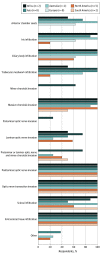
Results: Among the 52 survey recipients, the results are based on the responses from 27 individuals (52%) from 24 different retinoblastoma practices across 16 countries in 6 continents. The following were considered to be high-risk features: postlaminar optic nerve infiltration (27 [100%]), involvement of optic nerve transection (27 [100%]), extrascleral tissue infiltration (27 [100%]), massive (≥3 mm) choroidal invasion (25 [93%]), microscopic scleral infiltration (23 [85%]), ciliary body infiltration (20 [74%]), trabecular meshwork invasion (18 [67%]), iris infiltration (17 [63%]), anterior chamber seeds (14 [52%]), laminar optic nerve infiltration (13 [48%]), combination of prelaminar and laminar optic nerve infiltration and minor choroidal invasion (11 [41%]), minor (<3 mm) choroidal invasion (5 [19%]), and prelaminar optic nerve infiltration (2 [7%]). The other histopathologic features considered high risk included Schlemm canal invasion (4 [15%]) and severe anaplasia (1 [4%]). Four respondents (15%) said that the presence of more than 1 high-risk feature, especially a combination of massive peripapillary choroidal invasion and postlaminar optic nerve infiltration, should be considered very high risk for metastasis.
Conclusions and relevance: Responses to this nonvalidated survey conducted in 2020-2021 showed little uniformity in the definition of high-risk retinoblastoma. Postlaminar optic nerve infiltration, involvement of optic nerve transection, and extrascleral tumor extension were the only features uniformly considered as high risk for metastasis across all oncology practices. These findings suggest that the relevance about their value in the current scenario with advanced disease being treated conservatively needs further evaluation; there is also a need to arrive at consensus definitions and conduct prospective multicenter studies to understand their relevance.
Conflict of interest statement
Figures
References
Publication types
MeSH terms
LinkOut - more resources
Full Text Sources
Medical


