Magnetic resonance imaging predicted the therapeutic response of patients with spinal cerebrospinal fluid leakage undergoing targeted epidural blood patch
- PMID: 34762485
- PMCID: PMC8722244
- DOI: 10.1259/bjr.20210841
Magnetic resonance imaging predicted the therapeutic response of patients with spinal cerebrospinal fluid leakage undergoing targeted epidural blood patch
Abstract
Objective: Most patients with spinal cerebrospinal fluid (CSF) leakage require an epidural blood patch (EBP); however, the response to treatment is varied. This study aimed to compare the MRI findings at follow-up between EBP effective and non-effective groups and to identify imaging findings that predict EBP treatment failure.
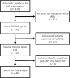
Methods: We retrospectively reviewed 48 patients who received EBP treatment for spinal CSF leakage. These patients were stratified into two groups: EBP effective (n = 27) and EBP non-effective (n = 21) using the results of the 3 month MRI as the end point.
Results: Compared to the EBP non-effective group, the patients in the EBP effective group had a lower spinal CSF leakage number (2.67 vs 12.48; p = 0.001), lower spinal epidural fluid accumulation levels (3.00 vs 7.48; p = 0.004), brain descend (11.11% vs 38.10%; p = 0.027), pituitary hyperemia (18.52% vs 57.14%; p = 0.007), and decreased likelihood of ≥three numbers of spinal CSF leakage (25.93% vs 90.48%; p = 0.001) in the post-EBP MRI. Clinical non-responsiveness (OR: 57.84; 95% CI: 3.47-972.54; p = 0.005) and ≥three numbers of spinal CSF leakage (OR: 15.13; 95% CI: 1.45-159.06; p = 0.023) were associated with EBP failure. Between these variables, ≥three numbers of spinal CSF leakage identified using the post-EBP MRI demonstrated greater sensitivity in predicting EBP failure compared to clinical non-responsiveness (90.48% vs 61.9%).
Conclusion: The number of spinal CSF leakage identified using the post-EBP MRI with a cut-off value of three is an effective predictor of EBP failure.
Advances in knowledge: Compared to clinical responsiveness, the post-EBP MRI provided a more objective approach to predict the effectiveness of EBP treatment in patients with spinal CSF leakage.
Conflict of interest statement
Figures


Similar articles
-
Risk factors for nonresponsive hydration in patients with spinal cerebrospinal fluid leakage.BMC Neurol. 2021 Nov 3;21(1):427. doi: 10.1186/s12883-021-02464-6. BMC Neurol. 2021. PMID: 34732159 Free PMC article.
-
Epidural Blood Patching in Spontaneous Intracranial Hypotension-Do we Really Seal the Leak?Clin Neuroradiol. 2023 Mar;33(1):211-218. doi: 10.1007/s00062-022-01205-7. Epub 2022 Aug 26. Clin Neuroradiol. 2023. PMID: 36028627 Free PMC article.
-
Efficacy and safety of cervicothoracic epidural blood patch for patients with spontaneous intracranial hypotension.Pain Pract. 2022 Jul;22(6):586-591. doi: 10.1111/papr.13126. Epub 2022 May 27. Pain Pract. 2022. PMID: 35585760
-
Surgical Management of Symptomatic Boxing-Induced Spinal Cerebrospinal Fluid Leak After Failed Epidural Blood Patch.World Neurosurg. 2020 Jul;139:478-482. doi: 10.1016/j.wneu.2020.04.194. Epub 2020 May 4. World Neurosurg. 2020. PMID: 32376374 Review.
-
Cervical epidural blood patch--A literature review.Pain Med. 2015 Oct;16(10):1897-904. doi: 10.1111/pme.12793. Epub 2015 Jun 30. Pain Med. 2015. PMID: 26122010 Review.
Cited by
-
Prediction of Target Epidural Blood Patch Treatment Efficacy in Spontaneous Intracranial Hypotension Using Follow-Up MRI.Diagnostics (Basel). 2022 May 6;12(5):1158. doi: 10.3390/diagnostics12051158. Diagnostics (Basel). 2022. PMID: 35626313 Free PMC article.
-
Quantitative Measurement of Spinal Cerebrospinal Fluid by Cascade Artificial Intelligence Models in Patients with Spontaneous Intracranial Hypotension.Biomedicines. 2022 Aug 22;10(8):2049. doi: 10.3390/biomedicines10082049. Biomedicines. 2022. PMID: 36009595 Free PMC article.
References
Publication types
MeSH terms
LinkOut - more resources
Full Text Sources
Medical

