. 2021 Dec 10;374(6573):eabm4805.
doi: 10.1126/science.abm4805. Epub 2021 Dec 10.

Computed structures of core eukaryotic protein complexes

Affiliations

Computed structures of core eukaryotic protein complexes

Ian R Humphreys et al. Science. .

Abstract

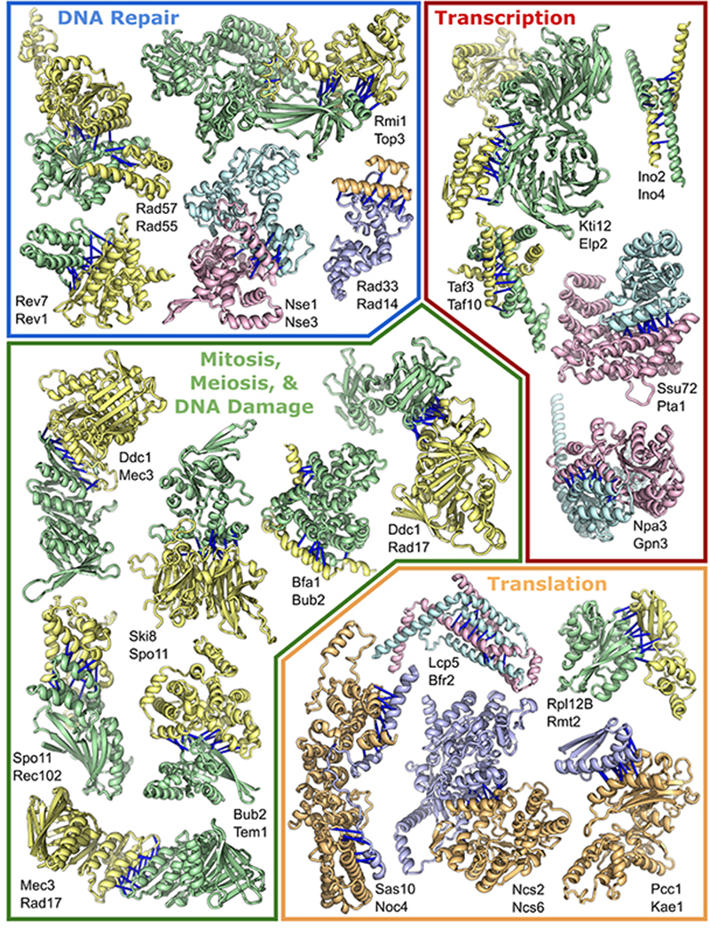
Protein-protein interactions play critical roles in biology, but the structures of many eukaryotic protein complexes are unknown, and there are likely many interactions not yet identified. We take advantage of advances in proteome-wide amino acid coevolution analysis and deep-learning–based structure modeling to systematically identify and build accurate models of core eukaryotic protein complexes within the Saccharomyces cerevisiae proteome. We use a combination of RoseTTAFold and AlphaFold to screen through paired multiple sequence alignments for 8.3 million pairs of yeast proteins, identify 1505 likely to interact, and build structure models for 106 previously unidentified assemblies and 806 that have not been structurally characterized. These complexes, which have as many as five subunits, play roles in almost all key processes in eukaryotic cells and provide broad insights into biological function.


Conflict of interest statement

Competing interests: Authors declare that they have no competing interests.

Figures

Figure 1
Figure 1. Evaluation of protein interaction and structure prediction accuracy.
(A) The PPI screen pipeline. (B) Performance (precision at different levels of recall) of different methods in picking out gold standard PPIs from the set of 4.3 million pMSAs (Precision: number of true positives above a cutoff divided by the total number of pairs above this cutoff; recall: number of true positives above cutoff divided by the total number of true positives (gold standard PPIs). Pairs were ranked by the top coevolution score or contact probability between residue pairs. DCA: Direct coupling analysis. RF2t: top contact probability between residues of two proteins by RF 2-track model. RF2t++, optimized RF2t (see methods). RF2t++ predictions better than the cutoff shown in vertical black line (RF2t++L in Fig. 1C) were processed with AF; recall of gold standard PPIs at this cutoff is 29%; and precision is 23%. RF2t++ results with a more stringent cutoff (red vertical line) are also shown in Fig 1C (RF2t++H). (C) AF contact probability ranking of complexes selected by RF2t++ in panel (B); complexes with scores above the horizontal black line were selected for further analysis. (D) Number of high scoring (top contact probability > 0.67) AF predictions in PPI sets from different sources. (E) Distribution of percent of AF predicted inter-protein contacts with predicted error < 8Å found in contact (< 8Å) in closely-related experimental structures.
Figure 2
Figure 2. Protein complexes involved in transcription, translation, and DNA repair.
Top predicted residue-residue contacts are indicated with bars. Pair color indicates the method of identification: pairs from the “pooled experimental sets → AF” screen are yellow and green, pairs from the “de novo RF → AF” screen are in blue and light-orange; and pairs present in both datasets are teal and pink. Full names of these proteins are in table S2.
Figure 3
Figure 3. Protein complexes involved in molecule transport, membrane translocation, and mitochondria.
Bars and coloring as in Fig 2. Full names for proteins are in table S3. Membrane spanning regions are annotated on Vtc1-Vtc4 and Sed5-Sft2. Top left: model of Vtc1-Vtc4 complex, with superimposed crystal structure (PDB: 3G3Q, Chain A) of the VTC4 (bright yellow) with phosphate bound (red balls).
Figure 4
Figure 4. Protein complexes involved in metabolism, GPI anchor biosynthesis or including a protein of unknown function.
Coloring is as in Fig. 2-3. Proteins annotated in the Uniprot database as uncharacterized proteins are denoted with an asterisk. Full names for these proteins are in table S4.
Figure 5
Figure 5. Higher order protein complexes.
(A) Top predicted residue-residue contacts for trimers are indicated with bars. Bar color corresponds to the interacting protein pair; protein 1:2 are blue, 1:3 are red, 2:3 are purple. Full names of each protein within the complex are in table S5. (B) Model of Rad55-Rad57-Rad51 and cartoon depiction of placement of this complex in the larger Rad51 filament. Additional information in fig. S18. (C) GARP complex model constructed by predicting structure of central hetero-oligomeric helical bundle, and superimposing models of individual components onto this. 2D class average of GARP complex with minor adaptation (77); reprinted by permission from Springer Nature Customer Service Center GmbH: Springer Nature, Nature Structural and Molecular Biology, CATCHR, HOPS and CORVET tethering complexes share a similar architecture, H-T Chou, D. Dukovski, M.G. Chambers, K.M. Reinisch, and T. Walz, 2016). Alternative GARP models are in fig. S24. (D) Rad33-Rad14 complex model superimposed onto previously determined TFIIH/Rad4-Rad23-Rad33 complex structure (7k04). See fig. S19 for additional details. (E) GPI-T pentamer model highlighting a possible peptide substrate recognition channel adjacent to the catalytic dyad. See fig. S27 for additional details.

Comment in

References

    1. Ito T, Chiba T, Ozawa R, Yoshida M, Hattori M, Sakaki Y. A comprehensive two-hybrid analysis to explore the yeast protein interactome. Proc Natl Acad Sci U S A. 2001;98:4569–4574. - PMC - PubMed
    1. Collins SR, Kemmeren P, Zhao X-C, Greenblatt JF, Spencer F, Holstege FCP, Weissman JS, Krogan NJ. Toward a comprehensive atlas of the physical interactome of Saccharomyces cerevisiae. Mol Cell Proteomics. 2007;6:439–450. - PubMed
    1. Reguly T, Breitkreutz A, Boucher L, Breitkreutz B-J, Hon GC, Myers CL, Parsons A, Friesen H, Oughtred R, Tong A, Stark C, et al. Comprehensive curation and analysis of global interaction networks in Saccharomyces cerevisiae. J Biol. 2006;5:11. - PMC - PubMed
    1. Uetz P, Giot L, Cagney G, Mansfield TA, Judson RS, Knight JR, Lockshon D, Narayan V, Srinivasan M, Pochart P, Qureshi-Emili A, et al. A comprehensive analysis of protein-protein interactions in Saccharomyces cerevisiae. Nature. 2000;403:623–627. - PubMed
    1. Yu H, Braun P, Yildirim MA, Lemmens I, Venkatesan K, Sahalie J, Hirozane-Kishikawa T, Gebreab F, Li N, Simonis N, Hao T, et al. High-quality binary protein interaction map of the yeast interactome network. Science. 2008;322:104–110. - PMC - PubMed

Publication types

MeSH terms