. 2021 Nov 11;17(11):e1010034.
doi: 10.1371/journal.ppat.1010034. eCollection 2021 Nov.

Siglec-9 defines and restrains a natural killer subpopulation highly cytotoxic to HIV-infected cells

Affiliations

Siglec-9 defines and restrains a natural killer subpopulation highly cytotoxic to HIV-infected cells

Opeyemi S Adeniji et al. PLoS Pathog. .

Abstract

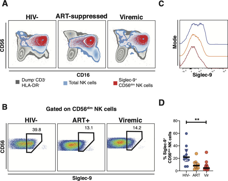
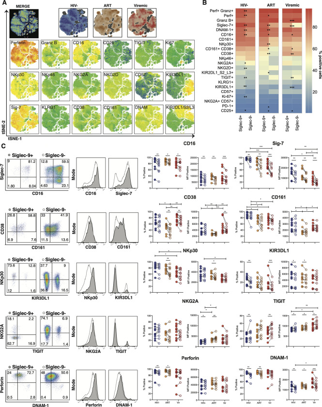
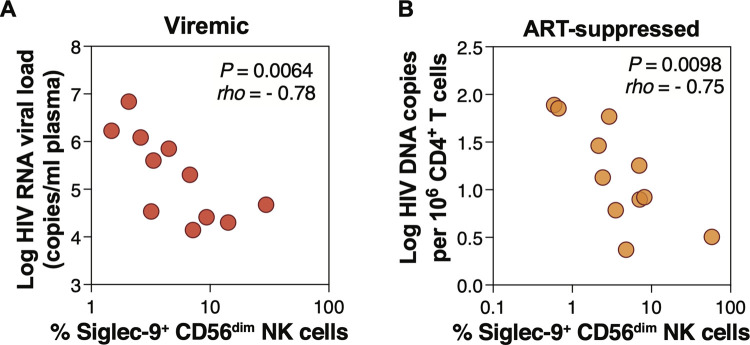
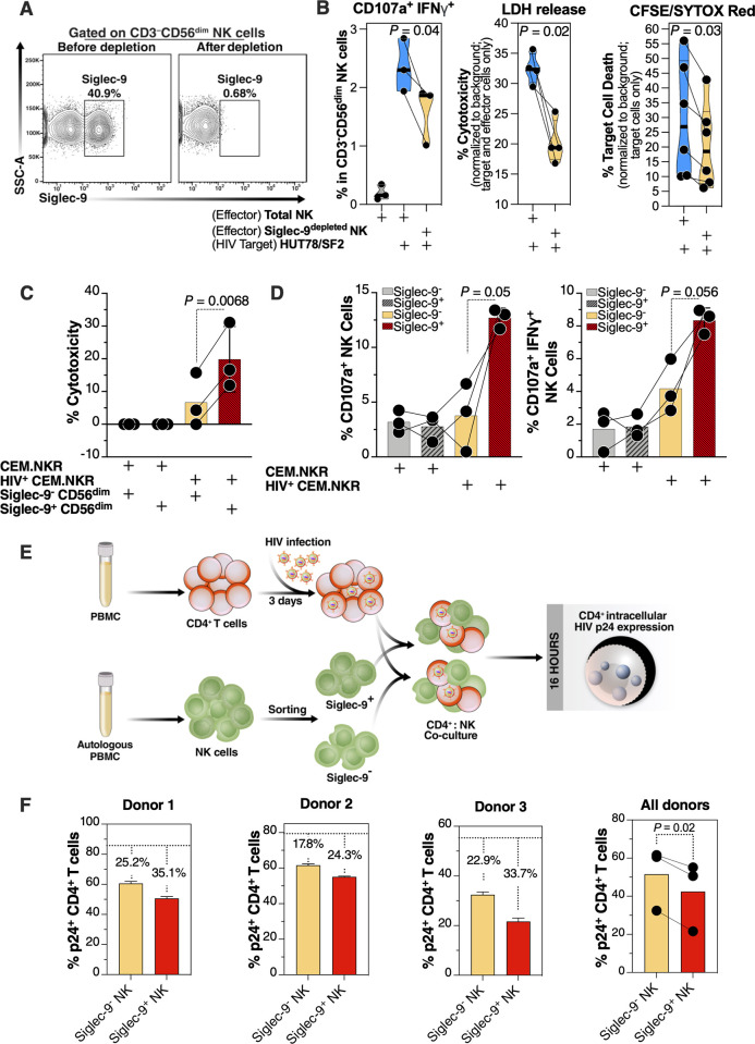
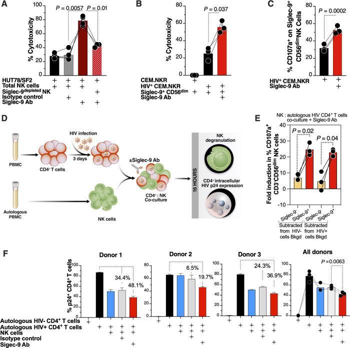
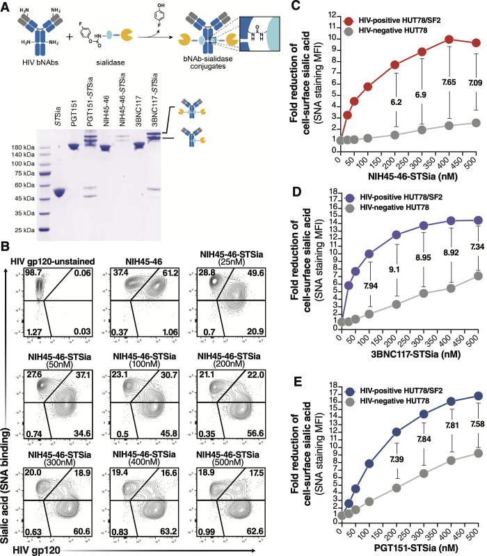
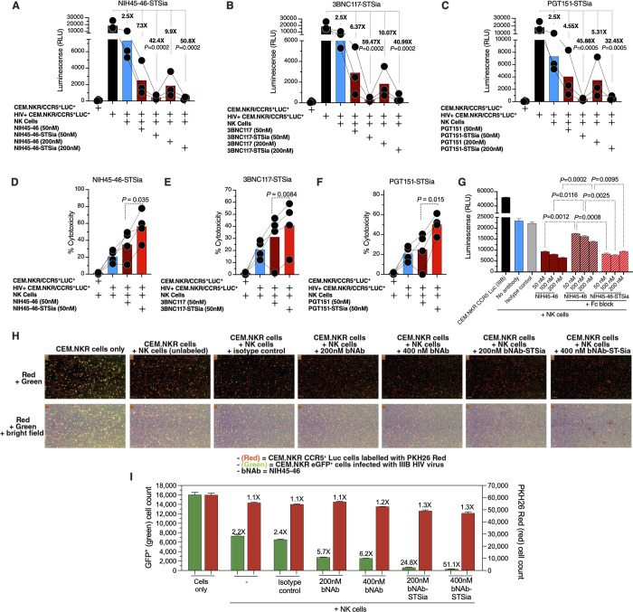
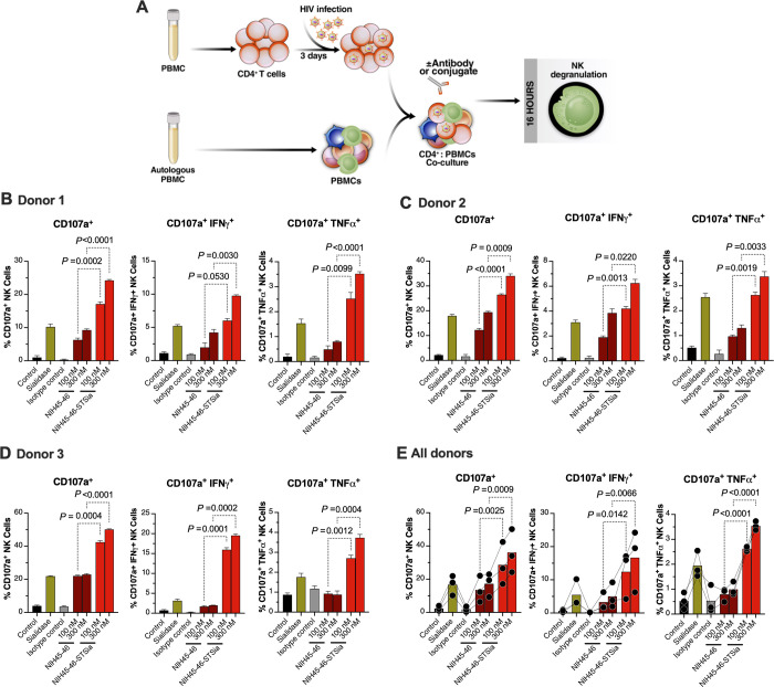
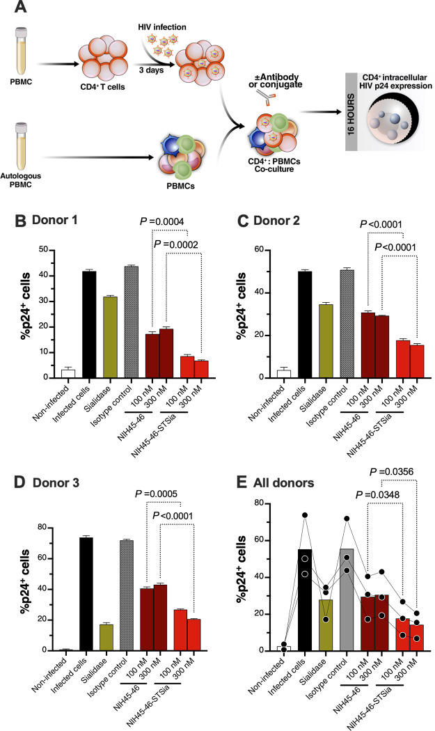
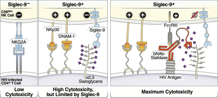
Siglec-9 is an MHC-independent inhibitory receptor expressed on a subset of natural killer (NK) cells. Siglec-9 restrains NK cytotoxicity by binding to sialoglycans (sialic acid-containing glycans) on target cells. Despite the importance of Siglec-9 interactions in tumor immune evasion, their role as an immune evasion mechanism during HIV infection has not been investigated. Using in vivo phenotypic analyses, we found that Siglec-9+ CD56dim NK cells, during HIV infection, exhibit an activated phenotype with higher expression of activating receptors and markers (NKp30, CD38, CD16, DNAM-1, perforin) and lower expression of the inhibitory receptor NKG2A, compared to Siglec-9- CD56dim NK cells. We also found that levels of Siglec-9+ CD56dim NK cells inversely correlate with viral load during viremic infection and CD4+ T cell-associated HIV DNA during suppressed infection. Using in vitro cytotoxicity assays, we confirmed that Siglec-9+ NK cells exhibit higher cytotoxicity towards HIV-infected cells compared to Siglec-9- NK cells. These data are consistent with the notion that Siglec-9+ NK cells are highly cytotoxic against HIV-infected cells. However, blocking Siglec-9 enhanced NK cells' ability to lyse HIV-infected cells, consistent with the known inhibitory function of the Siglec-9 molecule. Together, these data support a model in which the Siglec-9+ CD56dim NK subpopulation is highly cytotoxic against HIV-infected cells even whilst being restrained by the inhibitory effects of Siglec-9. To harness the cytotoxic capacity of the Siglec-9+ NK subpopulation, which is dampened by Siglec-9, we developed a proof-of-concept approach to selectively disrupt Siglec/sialoglycan interactions between NK and HIV-infected cells. We achieved this goal by conjugating Sialidase to several HIV broadly neutralizing antibodies. These conjugates selectively desialylated HIV-infected cells and enhanced NK cells' capacity to kill them. In summary, we identified a novel, glycan-based interaction that may contribute to HIV-infected cells' ability to evade NK immunosurveillance and developed an approach to break this interaction.


Conflict of interest statement

The authors have declared that no competing interests exist.

Figures

Fig 1
Fig 1. Expression of Siglec-9+ CD56dim NK cells during HIV infection.
(A) Overlay plots showing the distribution of Siglec-9+ CD56dim NK cells (red) compared with total NK cells (blue) and non-T cell lymphocytes (grey). (B-C) Representative plots showing the frequency (B) and expression (MFI overlay) (C) of Siglec-9 in total CD56dim NK cells in HIV- (blue line), HIV+ ART+ (orange line), and HIV+ viremic (red line) individuals. (D) Decreased frequency of Siglec-9+ CD56dim NK cells during HIV infection compared to HIV- controls. Lines in graphs indicate the median of the group. ** p<0.01. Mann-Whitney rank test was used to compare between groups. n = 10 HIV-negative controls, 11 HIV+ viremic, and 10 HIV+ on suppressive ART.
Fig 2
Fig 2. The phenotype of Siglec-9+ CD56dim NK cells.
(A) Global t-SNE visualization of Siglec-9+ CD56dim NK cells for all individuals pooled, with Siglec-9+ CD56dim NK cells from HIV-, HIV+ ART+, and HIV+ viremic individuals concatenated and overlayed (dimensionality reduction performed from 234,000 cells in 21 dimensions, 10,000 iterations, excluding parameters used to define the population: time, FSC, SSC, viability, CD14, CD19, CD3, and Siglec-9). Bottom: t-SNE projections of the 18 indicated proteins expression. (B) Heatmaps showing the percentages of Siglec-9+ and Siglec-9- CD56dim NK cells expressing the indicated activation and inhibitory markers in HIV-, HIV+ ART+, and HIV+ viremic individuals. (C) Comparative analyses of frequency (% positive) and expression (MFI of the positive population) of CD16, Siglec-7, CD38 CD161, NKp30, KIR3DL1, NKG2A, TIGIT, Perforin, and DNAM-1 on Siglec-9+ vs. Siglec-9- CD56dim NK cells. Left: Representative flow plots and histograms from HIV+ ART-suppressed donors are shown. Numbers inside the plots represent the gated percentage within the parent population. Mann-Whitney rank test was used to compare between groups. Paired Wilcoxson test was used to compare Siglec-9+ and Siglec-9- within each group. ***p<0.001, ** p<0.01, *p<0.05. n = 10 HIV-negative controls, 11 HIV+ viremic, and 10 HIV+ on suppressive ART.
Fig 3
Fig 3. Frequency of Siglec-9+ CD56dim NK cells correlates with viral load during viremic HIV infection and levels of CD4+ T cell-associated HIV DNA during ART-suppressed HIV infection.
(A) Spearman correlation between the frequency of Siglec-9+ CD56dim NK cells and HIV plasma viral load during viremic HIV infection. n = 11. (D) Spearman correlation between the frequency of Siglec-9+ CD56dim NK cells and cell-associated HIV DNA copies per million CD4+ T cells during ART-suppressed HIV infection. n = 11.
Fig 4
Fig 4. Siglec-9+ CD56dim NK cells exhibit higher cytotoxicity towards HIV+ cells compared to Siglec-9- CD56dim NK cells.
(A) A representative example of depletion of Siglec-9+ NK cells. (B) Siglec-9depleted NK cells exhibit lower cytotoxicity towards HIV-infected HUT78/SF2 targets compared to total NK cells. Cytotoxicity was assessed using NK degranulation, left panel (n = 3 donors; E:T = 4:1), LDH release, middle panel (n = 4 donors; E:T = 10:1), and CFSE/SYTOX Red assay, right panel (n = 6 donors; E:T = 10:1). NK degranulation measured as CD107a+ IFNγ+. Assays from each donor were performed in 2–4 replicates, and the average of these replicates per donor was used for statistical analyses. Statistical analyses were performed using paired t-tests. (C-D) FACS sorted Siglec-9+ CD56dim NK cells exhibit higher cytotoxicity towards HIV+ CEM.NKR targets compared to Siglec-9- CD56dim NK cells. (C) Cytotoxicity was assessed using LDH release assay (n = 3 donors, E:T = 10:1). (D) Analysis of NK degranulation (n = 3 donors; E:T = 4:1) was made on total NK cells gated on Siglec-9+ or Siglec-9- CD56dim NK cell subsets. Siglec-9+ = Siglec-9+ CD56dim NK cells and Siglec-9- = Siglec-9- CD56dim NK cells. Statistical analyses were performed using paired t-tests. (E) A schematic representation of the workflow to evaluate the cytotoxic potential of Siglec-9+ and Siglec-9- CD56dim NK cells against autologous HIV-infected CD4+ T cells. CD4+ T cells were isolated from fresh PBMC and exposed to HIV-1 IIIB for 72 h. On the third day, effector NK cells were isolated from PBMC of the same donor, FACS sorted, and co-cultured with autologous HIV-infected CD4+ T cells for 16 h. Following overnight incubation, the mixtures were stained for live/dead viability, CD3, and intracellular p24. (F) Data from the experimental design shown in (D). Dashed lines denote the percentage of p24+ cells in control HIV-infected CD4+ T cells cultured without effector cells. Percentages are percent reduction from dashed line. Assay from each donor was performed in triplicate (E:T = 10:1; n = 3 donors). Statistical analysis was performed using paired t-test.
Fig 5
Fig 5. The cytotoxicity of Siglec-9+ CD56dim NK cells towards HIV+ cells is being restrained by the inhibitory nature of the Siglec-9 molecule.
(A) HIV-infected HUT78/SF2 cells were used as targets, and total or Siglec9depleted NK cells from HIV-negative donors were used as effectors in the presence/absence of isotype control or Siglec-9 blocking Ab. Cytotoxicity was assessed by LDH release assay (E:T = 10:1). n = 4 donors (the last condition was performed on n = 3); assays from each donor were performed in 2–4 replicates, and the average of these replicates was used for statistical analysis using paired t-tests. (B-C) Blocking Siglec-9 enhanced the ability of Siglec-9+ CD56dim NK cells to target HIV-infected CEM.NKR cells. Cytotoxicity was assessed by (B) the LDH release assay (E:T = 10:1) or (C) NK degranulation (E:T = 4:1). Analysis of NK degranulation was made on total NK cells gated on Siglec-9+ or Siglec-9- CD56dim NK cell subsets. n = 3 donors; assays from each donor were performed in 2–4 replicates, and the average was used for statistical analysis using paired t-tests. (D) A schematic representation of the workflow to evaluate effector NK degranulation and cytotoxic potential against autologous HIV-infected CD4+ T cells in the presence or absence of Siglec-9 Ab. CD4+ T cells were isolated from fresh PBMC and exposed to HIV-1 IIIB for 72 h. On the third day, effector NK cells were isolated from PBMC of the same donor and co-cultured with autologous HIV-infected CD4+ T cells in the presence or absence of Siglec-9 Ab for 16 h. Both NK degranulation and intracellular p24 expression were evaluated by flow cytometry. (E) NK degranulation (CD107a expression) from the experiment described in (D). Assay from each donor was performed in triplicate (E:T = 2.5:1; n = 3 donors). Bkgd = background. Statistical analysis was performed using Paired t-tests. (F) Intracellular p24 expression from the experiment described in (D). Assay from each donor was performed in triplicate wells (E:T = 10:1; n = 3 donors). Percentages are percent reduction from the HIV-infected cells only condition. Statistical analysis was performed using paired ANOVA with post-hoc Holm-Sidak method (to correct for multiple comparisons).
Fig 6
Fig 6. bNAb-Sialidase conjugates selectively target HIV+ cells for desialylation.
(A) Top: preparation of site-specifically labeled HIV bNAb-Sialidase (bNAb-STSia) conjugates. Antibody-binding peptide (light blue) genetically fused with Sialidase (yellow) is conjugated to bNAb using pClick. pClick enables a site-specific conjugation between the antibody-binding peptide with payload and Lys337 of antibodies. Bottom: SDS-PAGE analysis with non-reducing buffer of bNAb-Sia conjugates. The two new bands above 180 kDa are consistent with the formation of the mono- or double-STSia antibody complex. (B-C) A mixture of HUT78 cells (HIV-negative) and HUT78/SF2 cells (HIV+) was treated with escalating doses of NIH45-46-STSia. HIV gp120 was measured by a secondary antibody to NIH45-46, and Sialic acid levels were measured as binding to SNA lectin. Representative flow plots (B). The fold reduction shows that sialic acid was reduced by >7 fold on HIV+ cells compared to HIV-negative cells (C). (D-E) Cells were treated as in B/C but using the 3BNC117-STSia conjugate (D) or the PGT151-STSia conjugate (E). STSia = Sialidase from Salmonella typhimurium.
Fig 7
Fig 7. bNAbs-STSia conjugates promote higher NK cytotoxicity against HIV+ cells compared to bNAbs alone.
(A-C) Killing assay using HIV-infected CEM-NKR CCR5+ Luc+ cells as targets and HIV-negative primary NK cells as effectors (n = 3 donors; assays from each donor were performed in 2–4 replicates, and the average of these replicates was used for analysis) at E:T ratio of 10:1. Luminescence was measured as a marker of intact (unkilled) HIV+ cells. (A) NIH45-46 and its conjugate. (B) 3BNC117 and its conjugate. (C) PGT151 and its conjugate. P-values were calculated using paired ANOVA with post-hoc Holm-Sidak method (comparing each condition against the HIV+ cells alone condition). (D-F) Killing assay using HIV-infected CEM-NKR CCR5+ Luc+ cells as targets and HIV-negative primary NK cells as effectors (n = 4 donors; assays from each donor were performed in 2–4 replicates, and the average of these replicates was used for analysis). Cytotoxicity was assessed by the LDH release assay at an E:T ratio of 10:1. (D) NIH45-46 and its conjugate. (E) 3BNC117 and its conjugate. (F) PGT151 and its conjugate. p-values were calculated using paired t-tests. (G) NK cells were treated with human Fc receptor blocking solution prior to co-incubation with HIV-infected CEM.NKR CCR5+ Luc+ cells. Luminescence was measured following 16 h incubation at a 10:1 E:T ratio. Unpaired ANOVA with post-hoc Dunnett T3 method (to correct for multiple comparisons) was used for statistical analysis between the indicated groups. (H) Effector NK cells were isolated from PBMC of an ART-suppressed HIV+ donor (ART09) and co-cultured with a mixture of HIV-uninfected PKH26-labeled CEM.NKR CCR5+ Luc+ (red cells) and HIV-infected CEM.NKR eGFP+ cells (green cells). Cell mixture was treated with NIH45-46, NIH45-46STSia, or isotype control. After 24 of co-culture, the Celigo image cytometer was used to directly visualize and count the number of PKH26-labeled (red) and GFP+ (green) target cells. The panel shows representative images from two independent experiments. This experiment was performed in quadruplicate at E:T 10:1. (I) Plot of the raw GFP+ green (HIV-infected) cell count (left y-axis) and red PKH26-labeled (HIV-uninfected) cell counts (right y-axis) from (H). The fold reduction compares the average of each condition to the cell-only condition.
Fig 8
Fig 8. NIH45-46-STSia induces NK degranulation towards autologous primary HIV-infected CD4+ T cells.
(A) A schematic representation of the workflow to evaluate effector NK degranulation against autologous HIV-infected CD4+ T cells in the presence of bNAb or bNAb-STSia conjugate. CD4+ T cells were isolated from fresh PBMC and exposed to HIV-1 IIIB for 72 h. On the third day, virus-infected CD4+ T cells were treated or not with Sialidase, NIH45-46, NIH45-46-STSia, or isotype-matched control antibody. PBMC from the same donor were co-cultured with autologous HIV-infected CD4+ T cells in the presence of CD107a antibody for 16 h. Following overnight incubation, the mixtures were stained with a cocktail of antibodies for CD3, CD56, IFN-γ, and TNF-α. Percent NK cell positive for CD107a, IFN-γ, and TNF-α expression was derived after gating on CD3-CD56dim NK cells. Control = data from the NK cells + HIV-infected CD4+ T cells condition. All other conditions contain NK cells + HIV-infected CD4+ T cells, in addition to the indicated reagent. (B) Data from donor 1. (C) Data from donor 2. (D) Data from donor 3. (E) Average data from all donors. Assay from each donor was performed in 4 replicate wells (E:T 10:1; n = 3 donors). Statistical analyses for panels B-D were performed using unpaired ANOVA with post-hoc Dunnett T3 method (to correct for multiple comparisons) comparing the indicated groups. Statistical analysis for panel E was performed using paired ANOVA with post-hoc Holm-Sidak method (comparing the indicated conditions).
Fig 9
Fig 9. NIH45-46-STSia induces PBMC cytotoxicity towards autologous primary HIV-infected CD4+ T cells.
(A) A schematic representation of the workflow to evaluate the cytotoxicity of PBMC against autologous HIV-infected CD4+ T cells in the presence of bNAb or bNAb-STSia conjugate. CD4+ T cells were isolated from fresh PBMC and exposed to HIV-1 IIIB for 72 h. On the third day, virus-infected CD4+ T cells were treated or not with Sialidase, NIH45-46, NIH45-46-STSia, or isotype-matched control antibody. PBMC from the same donor were co-cultured with autologous HIV-infected CD4+ T cells for 16 h. Following overnight incubation, the mixtures were stained for live/dead viability, CD3, CD8, and intracellular p24. Percent p24+ was derived after gating on CD3+, CD8- and live cells. (B) Data from donor 1. (C) Data from donor 2. (D) Data from donor 3. (E) Average data from all donors. Assay from each donor was performed in 4 replicate wells (E:T 100:1; n = 3 donors). Statistical analyses for panels B-D were performed using unpaired ANOVA with post-hoc Dunnett T3 method (to correct for multiple comparisons) comparing the indicated groups. Statistical analysis for panel E was performed using paired ANOVA with post-hoc Holm-Sidak method (comparing the indicated conditions).
Fig 10
Fig 10. Model of how HIV bNAb-Sialidase conjugates may increase the cytotoxicity of Siglec-9+ NK cells against HIV-infected cells.
Left two panels: The Siglec-9+ CD56dim NK subset has high cytolytic activity, possibly due to elevated expression of several NK activating receptors and reduced expression of the inhibitory NKG2A, compared to Siglec-9- CD56dim NK cells. However, Siglec-9 itself is an inhibitory receptor whose signaling restrains the cytolytic ability of these otherwise highly cytotoxic Siglec-9+ CD56dim NK cells by binding to Sialic acid attached to protein or lipid backbones on the surface of target cells. Right panel: Siglec/Sialic acid interactions are being pursued as an approach to enhance NK cell cytotoxicity against cancer using antibodies conjugated to Sialidase. We developed a similar proof-of-concept approach–conjugating Sialidase to HIV bNAbs–that could be used in conjunction with strategies that reactivate HIV latently-infected cells to enhance NK cells’ capacity to clear HIV+ cells.

References

    1. Chun TW, Finzi D, Margolick J, Chadwick K, Schwartz D, Siliciano RF. In vivo fate of HIV-1-infected T cells: quantitative analysis of the transition to stable latency. Nature medicine. 1995;1(12):1284–90. Epub 1995/12/01. doi: 10.1038/nm1295-1284 . - DOI - PubMed
    1. Wong JK, Hezareh M, Gunthard HF, Havlir DV, Ignacio CC, Spina CA, et al.. Recovery of replication-competent HIV despite prolonged suppression of plasma viremia. Science. 1997;278(5341):1291–5. doi: 10.1126/science.278.5341.1291 . - DOI - PubMed
    1. Finzi D, Hermankova M, Pierson T, Carruth LM, Buck C, Chaisson RE, et al.. Identification of a reservoir for HIV-1 in patients on highly active antiretroviral therapy. Science. 1997;278(5341):1295–300. doi: 10.1126/science.278.5341.1295 . - DOI - PubMed
    1. Chun TW, Carruth L, Finzi D, Shen X, DiGiuseppe JA, Taylor H, et al.. Quantification of latent tissue reservoirs and total body viral load in HIV-1 infection. Nature. 1997;387(6629):183–8. doi: 10.1038/387183a0 . - DOI - PubMed
    1. Chun TW, Stuyver L, Mizell SB, Ehler LA, Mican JA, Baseler M, et al.. Presence of an inducible HIV-1 latent reservoir during highly active antiretroviral therapy. Proc Natl Acad Sci U S A. 1997;94(24):13193–7. Epub 1997/12/16. doi: 10.1073/pnas.94.24.13193 ; PubMed Central PMCID: PMC24285. - DOI - PMC - PubMed

Publication types

MeSH terms