Review
. 2022 May;34(3):354-375.
doi: 10.1177/10406387211057469. Epub 2021 Nov 11.

Bacterial and viral enterocolitis in horses: a review

Affiliations
Review

Bacterial and viral enterocolitis in horses: a review

Francisco A Uzal et al. J Vet Diagn Invest. 2022 May.

Abstract

Enteritis, colitis, and enterocolitis are considered some of the most common causes of disease and death in horses. Determining the etiology of these conditions is challenging, among other reasons because different causes produce similar clinical signs and lesions, and also because some agents of colitis can be present in the intestine of normal animals. We review here the main bacterial and viral causes of enterocolitis of horses, including Salmonella spp., Clostridium perfringens type A NetF-positive, C. perfringens type C, Clostridioides difficile, Clostridium piliforme, Paeniclostridium sordellii, other clostridia, Rhodococcus equi, Neorickettsia risticii, Lawsonia intracellularis, equine rotavirus, and equine coronavirus. Diarrhea and colic are the hallmark clinical signs of colitis and enterocolitis, and the majority of these conditions are characterized by necrotizing changes in the mucosa of the small intestine, colon, cecum, or in a combination of these organs. The presumptive diagnosis is based on clinical, gross, and microscopic findings, and confirmed by detection of some of the agents and/or their toxins in the intestinal content or feces.

Keywords: colitis; enteritis; enterocolitis; horses; review.


Conflict of interest statement

Declaration of conflicting interests: The authors declared no potential conflicts of interest with respect to the research, authorship, and/or publication of this article.

Figures

Figures 1–2.
Figures 1–2.
Clinical manifestations of colitis in a horse. Figure 1. Hemorrhagic diarrhea. Figure 2. Ultrasound of a horse with colitis. Notice thickened and edematous colonic wall and liquid content (L). The dotted line between A and + indicates the thickness of the colonic wall (3.28 cm).
Figures 3–8.
Figures 3–8.
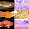
Equine enterocolitis of bacterial etiology. Figure 3. Colitis produced by Salmonella enterica serovar Typhimurium in a horse. The mucosa is hemorrhagic, necrotic, and covered by a fibrinous pseudomembrane. Photo courtesy of Dr. Francisco Carvallo. Figure 4. Colitis produced by S. enterica serovar Typhimurium in a horse. The mucosa is diffusely necrotic and there is transmural hemorrhage. H&E. Figure 5. Colitis produced by S. enterica serovar Typhimurium in a horse. There is a diffuse inflammatory infiltrate of the deep lamina propria. Inset: higher magnification showing large number of neutrophils in the lamina propria. H&E. Figure 6. Necrotic enteritis in a foal associated with NetF-positive Clostridium perfringens type A. Reproduced with permission from Mehdizadeh Gohari et al. Figure 7. Necrotic enteritis produced by C. perfringens type C in a neonatal foal. There is transmural hemorrhage and hemorrhagic content within the lumen. Figure 8. Necrotic enteritis produced by C. perfringens type C in a neonatal foal. There is severe and diffuse mucosal necrosis. Inset: higher magnification of the lamina propria and submucosal thrombosis. H&E.
Figures 9–14.
Figures 9–14.
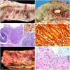
Equine enterocolitis of bacterial etiology. Figure 9. Colitis produced by Clostridioides difficile. The mucosa is diffusely hemorrhagic, and the content is liquid. Figure 10. Colitis produced by C. difficile. The mucosa is diffusely necrotic. Inset: higher magnification of volcano lesions. H&E. Figure 11. Colitis produced by Clostridium piliforme (Tyzzer disease). The mucosa is diffusely hemorrhagic, and the content is fluid. Figure 12. Colitis produced by C. piliforme (Tyzzer disease). Many filamentous silver-positive bacteria are in the cytoplasm of enterocytes and free in the intercellular spaces and lumen. Steiner. Figure 13. Necrotic enteritis produced by P. sordellii. There is severe and diffuse mucosal necrosis covered by a fibrous pseudomembrane and transmural hemorrhage. Figure 14. Necrotic enteritis produced by P. sordellii. There is severe and diffuse mucosal necrosis covered by a fibrous pseudomembrane, and transmural hemorrhage. Inset: higher magnification with clumps of intralesional rods. H&E.
Figures 15–20.
Figures 15–20.
Equine enterocolitis of bacterial or viral etiology. Figure 15. Hemorrhagic and ulcerative mucosa in colitis caused by Rhodococcus equi. Figure 16. Colonic mesenteric lymphadenitis caused by R. equi. The lymph node is enlarged and effaced by white, soft exudate. Figure 17. Submucosal granulomas in colitis caused by R. equi. H&E. Inset: higher magnification of the submucosal granulomas with large numbers of gram-positive coccobacilli in the cytoplasm of macrophages. Gram. Figure 18. Lymphoplasmacytic infiltration of the lamina propria in colitis caused by Neorickettsia risticii. Within macrophages and enterocytes are pinpoint, silver-positive, ~1-µm organisms compatible with N. risticii (arrows). Steiner. Figure 19. Diffusely hemorrhagic mucosa in enteritis caused by equine coronavirus. Photo courtesy of Dr. Federico Giannitti. Figure 20. Mucosal necrosis with crypt dilation in enteritis caused by equine coronavirus. H&E. Inset: immunohistochemistry for equine coronavirus. Photo courtesy of Dr. Federico Giannitti.

References

    1. Ackermann G, et al.. Isolation of Clostridium innocuum from cases of recurrent diarrhea in patients with prior Clostridium difficile associated diarrhea. Diagn Microbiol Infect Dis 2001;40:103–106. - PubMed
    1. Alberdi MP, et al.. Expression by Lawsonia intracellularis of type III secretion system components during infection. Vet Microbiol 2009;139:298–303. - PubMed
    1. Aldape MJ, et al.. Clostridium sordellii infection: epidemiology, clinical findings, and current perspectives on diagnosis and treatment. Clin Infect Dis 2006;43:1436–1446. - PubMed
    1. Arroyo LG, et al.. Experimental Clostridium difficile enterocolitis in foals. J Vet Intern Med 2004;18:734–738. - PubMed
    1. Arroyo LG, et al.. Duodenitis-proximal jejunitis in horses after experimental administration of Clostridium difficile toxins. J Vet Intern Med 2017;31:158–163. - PMC - PubMed