. 2021 Nov 11;12(1):6540.
doi: 10.1038/s41467-021-26750-6.

Targeting chondrocytes for arresting bony fusion in ankylosing spondylitis

Affiliations

Targeting chondrocytes for arresting bony fusion in ankylosing spondylitis

Fenli Shao et al. Nat Commun. .

Erratum in

Abstract

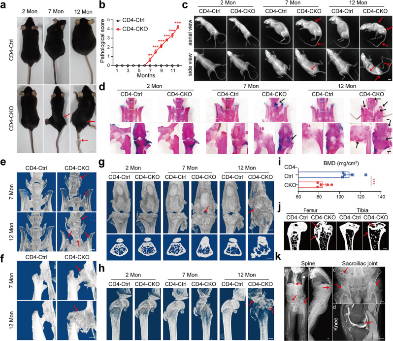
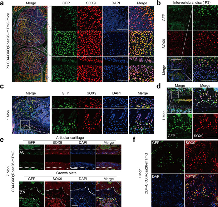
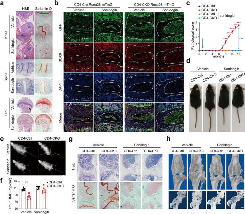
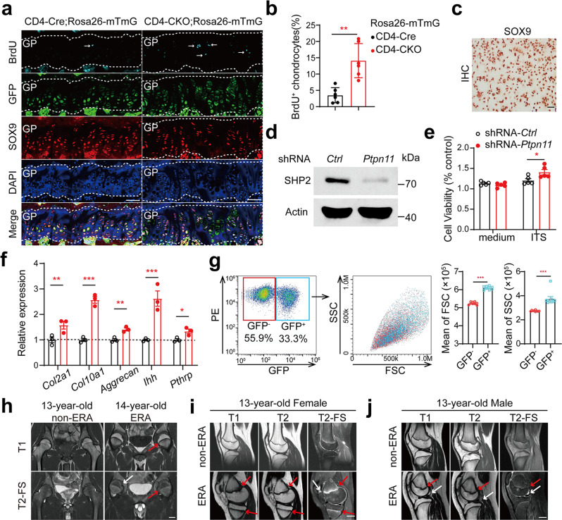
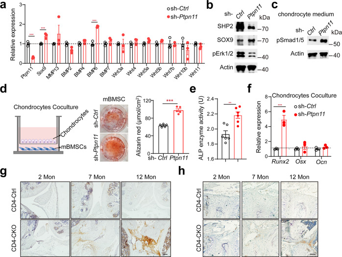
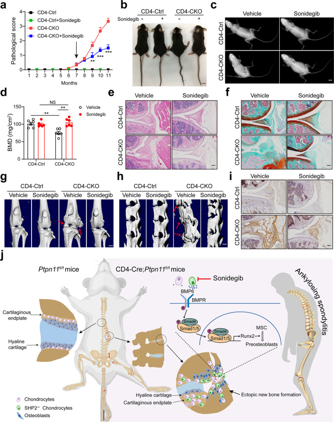
Bony fusion caused by pathological new bone formation manifests the clinical feature of ankylosing spondylitis (AS). However, the underlying mechanism remains elusive. Here we discovered spontaneous kyphosis, arthritis and bony fusion in mature CD4-Cre;Ptpn11f/f mice, which present the pathophysiological features of AS. A population of CD4-Cre-expressing proliferating chondrocytes was SHP2 deficient, which could differentiate into pre-hypertrophic and hypertrophic chondrocytes. Functionally, SHP2 deficiency in chondrocytes impeded the fusion of epiphyseal plate and promoted chondrogenesis in joint cavity and enthesis. Mechanistically, aberrant chondrocytes promoted ectopic new bone formation through BMP6/pSmad1/5 signaling. It is worth emphasizing that such pathological thickness of growth plates was evident in adolescent humans with enthesitis-related arthritis, which could progress to AS in adulthood. Targeting dysfunctional chondrogenesis with Smo inhibitor sonidegib significantly alleviated the AS-like bone disease in mice. These findings suggest that blockade of chondrogenesis by sonidegib would be a drug repurposing strategy for AS treatment.


Conflict of interest statement

Y.S. and Q.X. have a patent pending on use of sonidegib in ankylosing spondylitis. The other authors declare no competing interests.

Figures

Fig. 1
Fig. 1. Ptpn11 deletion in CD4-expressing cells causes age-related AS-like bone disease.
a Gross images of female mice. b Pathological scores (n = 10). c X‐ray images of female mice. Red arrows show kyphosis and joint ankylosis in mature and aged CD4-CKO mice. a, c Scale bars: 1 cm. d Skeleton staining results indicate increased Alcian blue staining (cartilage) in mature CD4-CKO mice and bone fusion in aged CD4-CKO mice. Black arrows show bone deformation. eh μ-CT radiographs show bone structure of sacroiliac joints, spines (e), hip joints, (f) and knee (g, h). Arrows show osteophytes and bony fusion in spine, hip joints and knee joints. i Femoral bone mineral density (BMD) (n = 5) and j Sagittal sections of femur and tibia show decreased BMD and loss of cortical bone and subchondral bone in aged female CD4-CKO mice. Arrows show bone loss. dh, j Scale bars: 1 mm. k Radiological images of patients with AS. Arrows indicate outward acetabular labrum, ankylosis of hip and sacroiliac joints and abnormity of epiphysis respectively. Scale bars: 2.5 cm. b, i Data are presented as mean ± SEM. b **p < 0.01, ***p < 0.001, i p-value = 0.0010, determined by two-tailed Student’s t-test. a, ch, j, k Data are representative of three independent biological replicates.
Fig. 2
Fig. 2. Ectopic new bone formation is accompanied by aberrant chondrocytes and inflammation in CD4-CKO mice.
a TRAP staining of distal femur stained. b Osteocalcin immunostaining images of tibia sections. Arrows indicate osteoclasts. c, d H&E and Safranin O-Fast Green (SOFG) analysis of knee joints. Black arrows show degenerated articular cartilage, and blue arrows show ectopic new bone formation in articular cavity and enthesis. e, f H&E and SOFG staining images of spine section. g H&E and SOFG staining images of hip joint section. ag Scale bars: 200 μm. c, d, g Black arrows show degenerated articular cartilage, and blue arrows show ectopic new bone formation in articular cavity and enthesis. Data are representative of three independent biological replicates. h, i SOFG staining images and measure of the epiphyseal plate. Scale bars: 50 μm. i Data are presented as mean ± SEM (n = 10). ***p < 0.001, determined by two-tailed Student’s t-test. j H&E staining images show inflammation, pannus formation, and proliferating synoviocytes in knee joints. Scale bars: 100 μm. Data are representative of five independent biological replicates.
Fig. 3
Fig. 3. Immune system abnormality is not the cause of AS-like bone deformation.
a Flow cytometry analysis of T cell subsets in inguinal lymph nodes. (n = 5). b Flow cytometry results of CD3+ T percentages in the spleen of 6-month-old recipient NCG mice (n = 4). c X‐ray images of 12-month-old recipient NCG. df Gross images (d), X‐ray images (e), and Femoral BMD (f) of Lck-CKO mice and Lck-Ctrl littermates. (n = 5). gk CD4-Ctrl (WT) and CD4-CKO (KO) mice at 4 months were lethally irradiated followed by transferring with bone marrow cells. Irradiated CD4-CKO mice transferred with WT bone marrow cells were labeled as WT-KO, and so on. Pathological scores (n = 10) (g), Gross images (h) and Radiographs (i) of 12-month-old chimeras. ce, hi Scale bars: 1 cm. j, k H&E and SOFG staining images of chimeras. Scale bars: 200 μm. j Arrows show bone deformation. k Arrows indicate chondrogenesis and ectopic new bone formation. a, b, f, g Data are presented as mean ± SEM. *p < 0.05. **p < 0.01, ***p < 0.001, determined by two-tailed Student’s t-test. ce, hk Data are representative of three independent biological replicates.
Fig. 4
Fig. 4. CD4-Cre mediates conditional deletion of SHP2 in chondrocytes.
Sagittal section of joints from CD4-CKO; Rosa26-mTmG mice were immunostained with marker protein GFP, transcriptional regulator SOX9 and nuclear counterstain DAPI. a, b Immunofluorescence staining images show SOX9 expression in all chondrocytes and GFP expression in hypertrophic chondrocytes in knee and spine section of P3 CD4-CKO; Rosa26-mTmG mice. a Blue dotted box shows cartilage mold, and green dotted box indicates hypertrophic zone of the growth plate. c, d Representative images of 1-month-old CD4-CKO; Rosa26-mTmG mice show GFP+ chondrocytes consisted in hypertrophic zone of femur and spine. e, f Immunofluorescence analysis of articular cartilage and growth plate (e) and chondroma (f) of the tibia in 7-month-old CD4-CKO; Rosa26-mTmG mice show GFP+ differentiated chondrocytes in the growth plate and articular cartilage of mature CD4-CKO;Rosa26-mTmG mice. e The area inside the two white dotted lines is articular cartilage (AC) (upper) and growth plate (GP) (under). af Scale bars, 100 μm. Data are representative of three independent experiments.
Fig. 5
Fig. 5. Premature fusion of the growth plate prevents AS-like bone disease in CD4-CKO mice.
Three-week-old CD4-CKO;Rosa26-mTmG mice and CD4-Cre;Rosa26-mTmG littermates were orally gavaged every other day with Smo inhibitor, sonidegib (50 mg/kg) for three times. a Sagittal joint sections of 2-month-old mice treated with sonidegib or not were stained with H&E and SOFG. Scale bars: 400 μm. b Spinal sections of 2-month-old mice were immunostained with marker protein GFP, transcriptional regulator SOX9 and nuclear counterstain DAPI. CE represents cartilaginous endplate between white dotted line. Scale bars: 200 μm. c Pathological scores of CD4-CKO mice treated with sonidegib or not (n = 10). df Gross images (d), X-ray radiographs (e), and femoral BMD (n = 6) (f) show bone lesions and decreased femoral BMD were merely occurred in CD4-CKO mice. Scale bars: 1 cm. g H&E and SOFG staining images. Scale bars: 400 μm. h μ-CT radiographs of 12-month-old mice show bone deformation and cartilage abnormalities in CD4-CKO mice without sonidegib treatment and merely smaller skeleton in sonidegib-treated CD4-CKO mice and CD4-Ctrl mice. Scale bars: 1 mm. c and f Data are presented as mean ± SEM. c **p < 0.01, ***p < 0.001, f **p-value = 0.0004, determined by two-tailed Student’s t-test. a, b, d, e, g, h Data are representative of three independent biological replicates.
Fig. 6
Fig. 6. Disorder of the growth plate occurs in CD4-CKO mice and juveniles with enthesitis-related arthritis.
a, b Mature mice were injected with 50 mg/kg BrdU intraperitoneally every 8 h for six times. Sections of knee joints were immunostained with BrdU, GFP, SOX9 and DAPI. (n = 6). GP: growth plate. The proportions of BrdU+SOX9+ chondrocytes in SOX9+ chondrocytes were measured. a Scale bars: 100 μm. c Mouse primary chondrocytes of C57BL/6 mice confirmed by SOX9 immunostaining. Scale bars: 100 μm. df Mouse chondrocytes transfected with shRNA-Ctrl or shRNA-Ptpn11 lentivirus respectively and the SHP2 protein level, cell proliferation activity and the expression of gene Col2a1, Col10a1 and Aggrecan were analyzed by Western blot (d), MTT activity (n = 5) (e) and RT-qPCR (n = 3) (f) respectively. g Flow cytometry analysis of Primary chondrocytes isolated from Ptpn11f/f;Rosa26-mTmG mice were transfected with half-dose Cre recombinase adenovirus. The mean FSC and SSC of GFP- and GFP+ chondrocytes were calculated. (n = 9). hj MRI with T1 weighed, T2 weighed and T2 weighed fat-saturated (T2-FS) sequence of patients with non-ERA or ERA. Hip joints (h), and knee joints (i, j) images show arthritis, bone deformation (white arrows), and thickener growth plate (red arrows) in joints of patients with ERA than those of age, sex-matched non-ERA patients. Scale bars: 2.5 cm. b, e, f, g Data are presented as mean ± SEM. *p < 0.05, **p < 0.01, ***p < 0.001, determined by two-tailed Student’s t-test. a, c, d, gj Data are representative of three independent biological replicates.
Fig. 7
Fig. 7. Aberrant chondrocytes promote ectopic new bone formation through BMP6/Smad1/5 signaling.
a RT-qPCR analyses of gene expression of chondrocytes after transfected with sh-Ctrl and sh-Ptpn11, respectively. (n = 3). b Western blot analyses of chondrocytes of (a). c western blot analysis of phosphorylation of Smad1/5 protein levels in mBMSC stimulated with chondrocytes supernatant for 30 min. d Alizarin Red staining and quantification of mBMSC cocultured with chondrocytes for 14 days. (n = 5). e, f ALP activity measurement (n = 6) (e) and RT-qPCR analyses (n = 3) (f) of osteogenic marker genes in mBMSC cocultured with chondrocytes for 7 days. e **p-value = 0.0020. (g and h) Immunostaining analysis of pSmad1/5 protein levels in knee joint (g) and intervertebral disc (h). Scale bars: 100 μm. a, d, e, f Data are presented as mean ± SEM. *p < 0.05, **p < 0.01, ***p < 0.001, determined by two-tailed Student’s t-test. bd, g, h Data are representative of three independent biological replicates.
Fig. 8
Fig. 8. Targeting chondrocyte retards AS progression in CD4-CKO mice.
7-month-old CD4-CKO mice and CD4-Cre littermates were orally gavaged with Smo inhibitor, sonidegib (100 mg/kg) for 4 months. a Pathological score (n = 6). b Gross images. c X‐ray images. (b and c) Scale bars: 1 cm. d Femur bone mineral density (BMD). (n = 6) e H&E staining of knee joints. f SOFG staining of knee joints. g and h μ-CT radiographs of knee joint (g) and spine (h) of 11-month-old mice. Arrows show ectopic new bone formation. Scale bars: 2.5 cm. i Immunohistochemical staining of pSmad1/5 protein levels in knee joint of 11-month-old mice. e, f, i Scale bars: 100 μm. a, d Data are presented as mean ± SEM. **p < 0.01, ***p < 0.001, determined by two-tailed Student’s t-test. b, c, ei Data are representative of three independent biological replicates. j The mechanism of AS-like bone disease in CD4-CKO mice.

References

    1. Sieper, J. & Poddubnyy, D. Axial spondyloarthritis. Lancet390, 73–84 (2017). - DOI - PubMed
    1. Taurog, J. D., Chhabra, A. & Colbert, R. A. Ankylosing spondylitis and axial spondyloarthritis. N. Engl. J. Med374, 2563–2574 (2016). - DOI - PubMed
    1. Cortes, A. et al. Identification of multiple risk variants for ankylosing spondylitis through high-density genotyping of immune-related loci. Nat. Genet45, 730–738 (2013). - DOI - PMC - PubMed
    1. de Vlam, K., Lories, R. J. & Luyten, F. P. Mechanisms of pathologic new bone formation. Curr. Rheumatol. Rep.8, 332–337 (2006). - DOI - PubMed
    1. Ward, M. M. et al. American college of rheumatology/spondylitis association of America/spondyloarthritis research and treatment network 2015 recommendations for the treatment of ankylosing spondylitis and nonradiographic axial spondyloarthritis. Arthritis Rheumatol.68, 282–298 (2016). - DOI - PMC - PubMed

Publication types