. 2021 Nov 11;11(1):22110.
doi: 10.1038/s41598-021-01475-0.

Chronic pathophysiological changes in the normal brain parenchyma caused by radiotherapy accelerate glioma progression

Affiliations

Chronic pathophysiological changes in the normal brain parenchyma caused by radiotherapy accelerate glioma progression

Yuichiro Tsuji et al. Sci Rep. .

Abstract

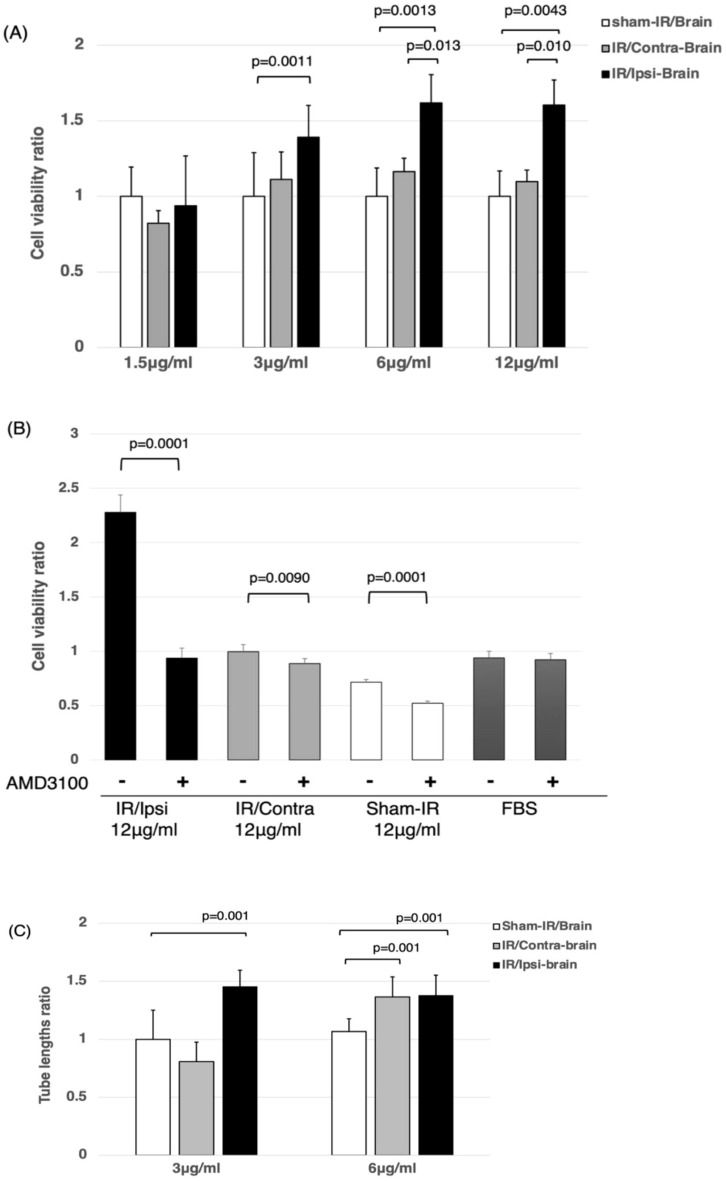
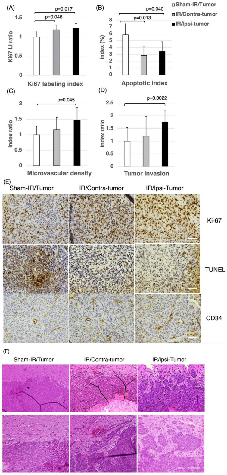
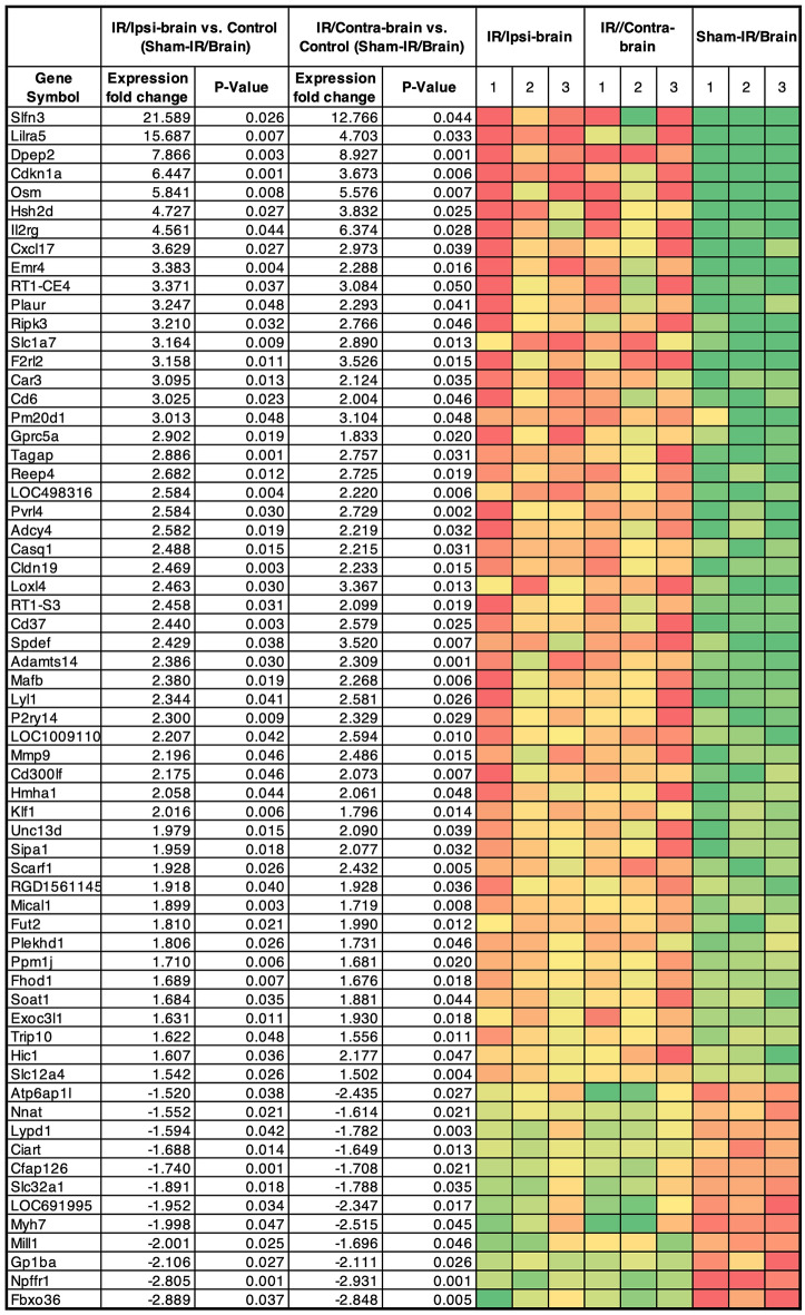
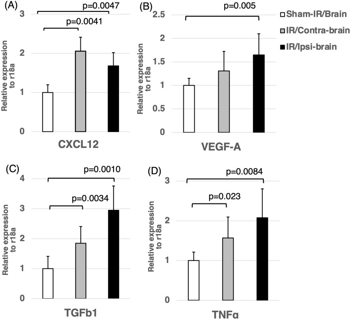
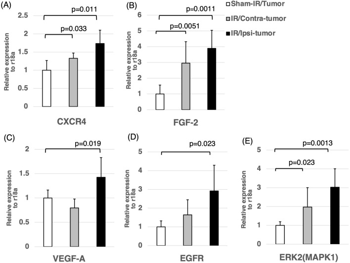
Radiation therapy is one of standard treatment for malignant glioma after surgery. The microenvironment after irradiation is considered not to be suitable for the survival of tumor cells (tumor bed effect). This study investigated whether the effect of changes in the microenvironment of parenchymal brain tissue caused by radiotherapy affect the recurrence and progression of glioma. 65-Gy irradiation had been applied to the right hemisphere of Fisher rats. After 3 months from irradiation, we extracted RNA and protein from the irradiated rat brain. To study effects of proteins extracted from the brains, we performed WST-8 assay and tube formation assay in vitro. Cytokine production were investigated for qPCR. Additionally, we transplanted glioma cell into the irradiated and sham animals and the median survival time of F98 transplanted rats was also examined in vivo. Immunohistochemical analyses and invasiveness of implanted tumor were evaluated. X-ray irradiation promoted the secretion of cytokines such as CXCL12, VEGF-A, TGF-β1 and TNFα from the irradiated brain. Proteins extracted from the irradiated brain promoted the proliferation and angiogenic activity of F98 glioma cells. Glioma cells implanted in the irradiated brains showed significantly high proliferation, angiogenesis and invasive ability, and the post-irradiation F98 tumor-implanted rats showed a shorter median survival time compared to the Sham-irradiation group. The current study suggests that the microenvironment around the brain tissue in the chronic phase after exposure to X-ray radiation becomes suitable for glioma cell growth and invasion.


Conflict of interest statement

The authors declare no competing interests.

Figures

Figure 1
Figure 1
(A) The cell-viability ratio under each protein concentration extracted from each group by WST-8 assay. Tissue proteins extracted from the IR/Ipsi-brain group contained significantly higher levels of growth factors compared to the Sham-IR/Brain group under the 3, 6, and 12 μg/ml concentrations of extracted protein. These results were expressed as mean ± SD and comparisons between groups were assessed by Student’s t-test. (B) The cell-viability ratio under 12 μg/ml concentrations of extracted protein from each group adding AMD3100(10 μM) by WST-8 assay. The cell-viability ratio adding AMD3100 in IR/Ipsi-brain, IR/Contra-brain, and Sham-IR/Brain groups was significantly inhibited. These results were expressed as mean ± SD and comparisons between groups were assessed by Student’s t-test. (C) Measurement of tube lengths by a tube-forming assay under each protein concentration extracted from each group. Tissue proteins extracted from the IR/Ipsi-brain group contained a significantly higher level of angiogenesis factors compared to those of the Sham-IR/Brain group. Under the protein concentration of 6 μg/ml, significantly longer tube formation was observed in the IR/Contra-brain group compared to the Sham-IR/Brain group. These results were expressed as mean ± SD and comparisons between groups were assessed by Student’s t-test.
Figure 2
Figure 2
(A,B) Survival curves of Fischer rats implanted with F98 glioma cells. Significantly shorter median survival times were achieved by the IR/Ipsi-tumor and IR/Contra-tumor rats compared to the Sham-IR/Tumor rats (IR/Ipsi-tumor and IR/Contra-tumor = 20.5 days vs Sham-IR/Tumor group = 22.5 days; p = 0.002 and p = 0.003, respectively). There was no significant difference in survival between the IR/Ipsi-tumor and IR/Contra-tumor groups (p = 0.8).
Figure 3
Figure 3
Changes in the biology of the transplanted tumors. (A) Ki-67 labeling index. (B) Apoptotic index. (C) Microvascular density (MVD) index. (D) Tumor invasion index. The tumors of the IR/Ipsi-tumor group exhibited significantly higher proliferation, angiogenesis abilities and invasiveness compared to those of the Sham-IR/Tumor group. Tumors of the IR/Ipsi-tumor and IR/Contra-tumor group showed significantly lower rates of apoptosis compared to the Sham-IR/Tumor group. These results were expressed as mean ± SD and comparisons between groups were assessed by Student’s t-test. (E) F98 glioma xenografts were stained immunohistochemically for Ki-67 (upper panels), TUNEL (middle panels) and CD34 (lower panels) at a magnification of × 400. Tumors of the IR/Ipsi-tumor and IR/Contra-tumor group showed a significant increase in proliferative index and a significant decrease in apoptotic index compared to Sham/IR-Tumor group. Tumors of the IR/Ipsi-tumor showed a significant increase in microvascular density index compared to Sham/IR-Tumor group. Scale bars = 50 μm. (F) Hematoxylin and eosin staining of F98 tumor showed tumor rim for sham-IR/Tumor (left panels), IR/Contra-tumor (middle panels) and IR/Ipsi-tumor (right panels). The upper panel shows the image at a magnification of × 80, and the area surrounded by the square shows the image at a magnification of × 200 (lower panel). Scale bars = 100 μm.
Figure 4
Figure 4
64 genes that showed significant expression changes of more than 1.5-fold or less than − 1.5-fold in both x-ray irradiated and non-irradiated hemisphere, compared to the sham-irradiated brain. Significance level is p < 0.05.
Figure 5
Figure 5
Expression of various cytokines in the IR/Ipsi-brain, IR/Contra-brain, and Sham-IR/Brain groups. The expressions of CXCL12, VEGF-A, TGF-β1 and TNFα in the IR/Ipsi-brain group were significantly higher compared to those in the Sham-IR/Brain group. The expressions of CXCL12, TGF-β1 and TNFα of the IR/Contra-brain group were significantly higher than those of the Sham-IR/Brain group. These results were expressed as mean ± SD and comparisons between groups were assessed by Student’s t-test.
Figure 6
Figure 6
Expression of various cytokines in the IR/Ipsi-tumor, IR/Contra-tumor, and Sham-IR/Tumor groups. The expressions of CXCR4, FGF-2, VEGF-A, EGFR and ERK2 in the IR/Ipsi-tumor group were significantly higher than those in the Sham-IR/Tumor group. The expressions of CXCR4, FGF-2 and ERK2 in the IR/Contra-tumor group were significantly higher compared to those in the Sham-IR/Tumor group. These results were expressed as mean ± SD and comparisons between groups were assessed by Student’s t-test.
Figure 7
Figure 7
(A) Schema of X-ray irradiation to the rat brain (created with BioRender.com). The whole body of rat was covered with a lead plate with a 1 cm square window, and only a part of the right cerebral hemisphere was irradiated with X-rays. (B) Treatment schema of the three groups of male Fisher rats for in vitro study (created with BioRender.com). X-ray: X-ray irradiation. IR/Ipsi-brain group: X-ray was irradiated only a part of the right cerebral hemisphere and the tissue of the right hemisphere was used. IR/Contra-brain group: X-ray was irradiated only a part of the right cerebral hemisphere and the tissue of the left hemisphere was used. Sham-IR/Brain group: no irradiation was performed and the tissue of the right hemisphere was used. (C) Treatment schema of the three groups of Fisher rats implanted tumor for in vivo study (created with BioRender.com). T: implanted tumor. IR/Ipsi-tumor group: F98 cells were transplanted to the right hemisphere after irradiation to the right hemisphere. IR/Contra-tumor group: F98 cells were transplanted to the left hemisphere after irradiation to the right hemisphere. Sham-IR/Tumor group: F98 cells were transplanted to the right hemisphere without irradiation. (D) Timeline describing the timing of irradiation, sacrifice and brain extraction in vitro assay. Three months after the X-ray irradiation, we extracted the tissue protein and RNA from rat brain. (E) Timeline describing the timing of irradiation, tumor implantation and sacrifice for in vivo assay. Three months after the irradiation, F98 glioma cells were implanted into the cerebral hemisphere of rats.

References

    1. Chinot OL, Wick W, Mason W, et al. Bevacizumab plus radiotherapy-temozolomide for newly diagnosed glioblastoma. N. Engl. J. Med. 2014;370:709–722. - PubMed
    1. Anderson AP. Postoperative irradiation of glioblastomas. Results in a randomized series. Acta. Radiol. Oncol. Radiat. Phys. Biol. 1978;17(6):475–484. - PubMed
    1. Walker MD, Green SB, Byar DP, et al. Randomized comparisons of radiotherapy and nitrosoureas for the treatment of malignant glioma after surgery. N. Engl. J. Med. 1980;303(23):1323–1329. - PubMed
    1. Gasper LE, Fisher BJ, Macdonald DR, et al. Supratentorial malignant glioma: patterns of recurrence and implications for external beam local treatment. Int. J. Radiat. Onco. Biol. Phys. 1992;24(1):55–57. - PubMed
    1. McDonald MW, Shu HK, Curran WJ, Jr, et al. Pattern of failure after limited margin radiotherapy and temozolomide for glioblastoma. Int. J. Radiat. Oncol. Biol. Phys. 2011;79(1):130–136. - PubMed

Publication types