. 2021 Oct 29;54(5):143-153.
doi: 10.1267/ahc.21-00012. Epub 2021 Oct 2.

ZMYM3 May Promote Cell Proliferation in Small Cell Lung Carcinoma

Affiliations

ZMYM3 May Promote Cell Proliferation in Small Cell Lung Carcinoma

Noritaka Kudo et al. Acta Histochem Cytochem. .

Abstract

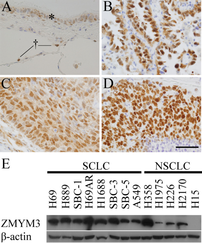
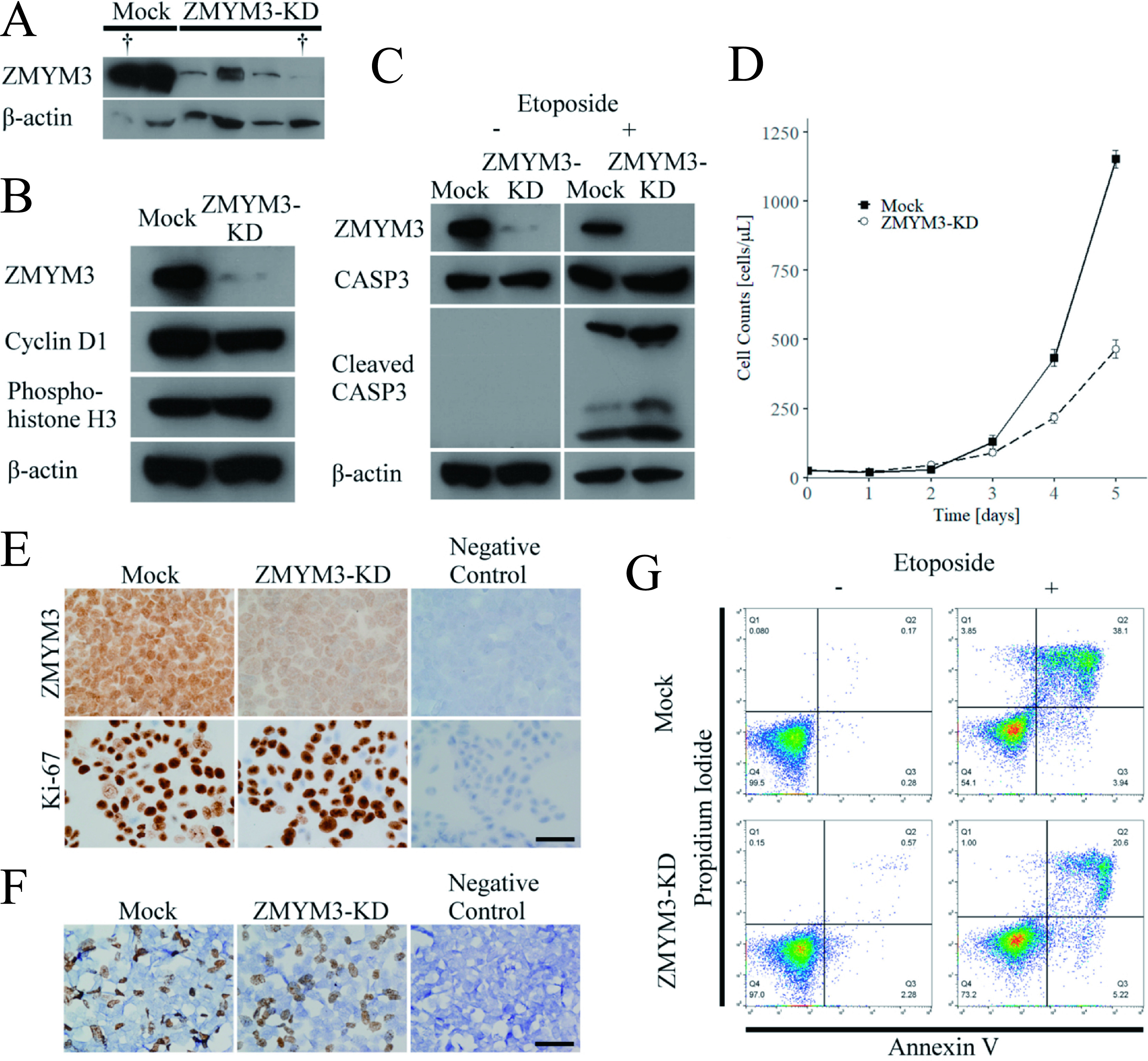
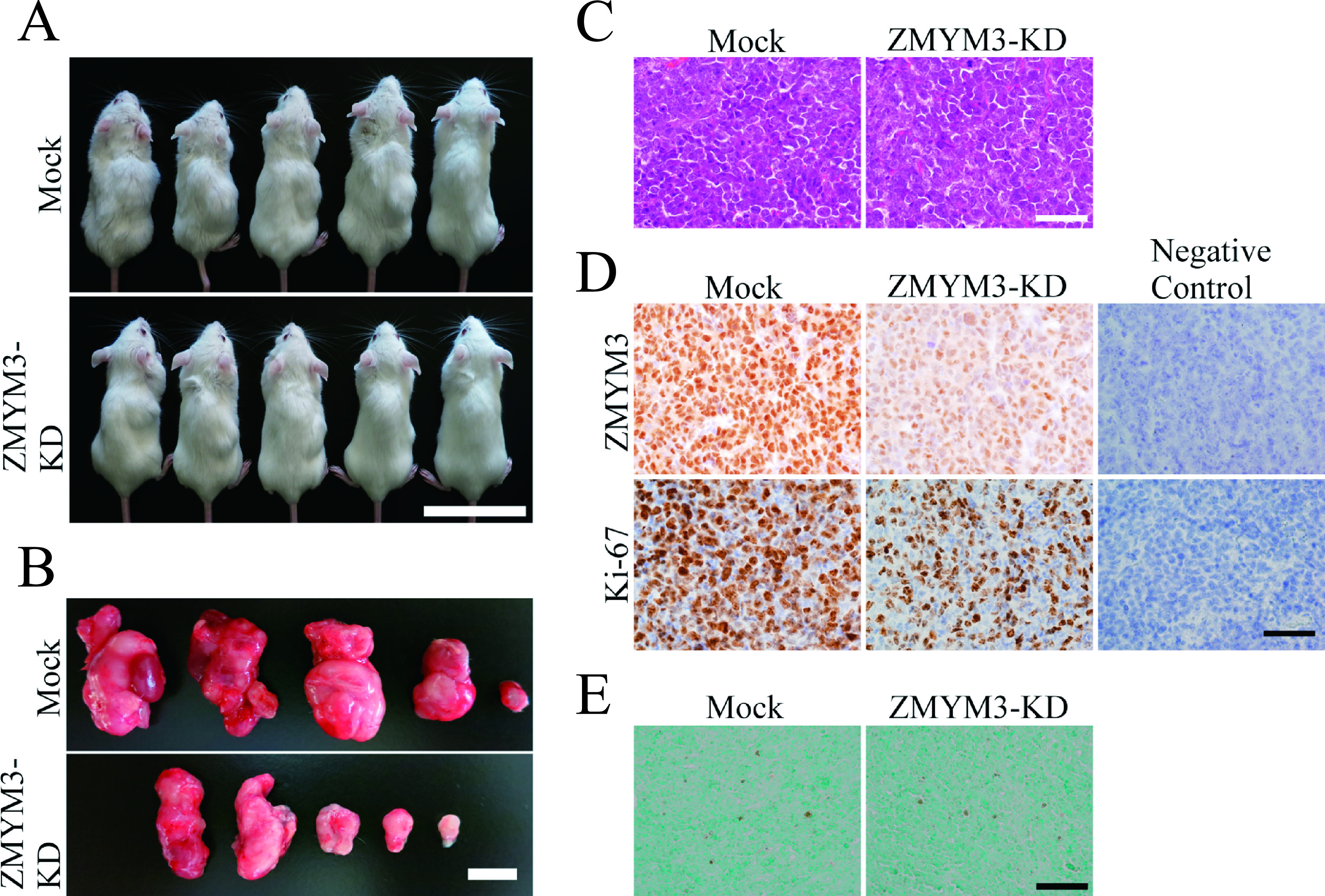
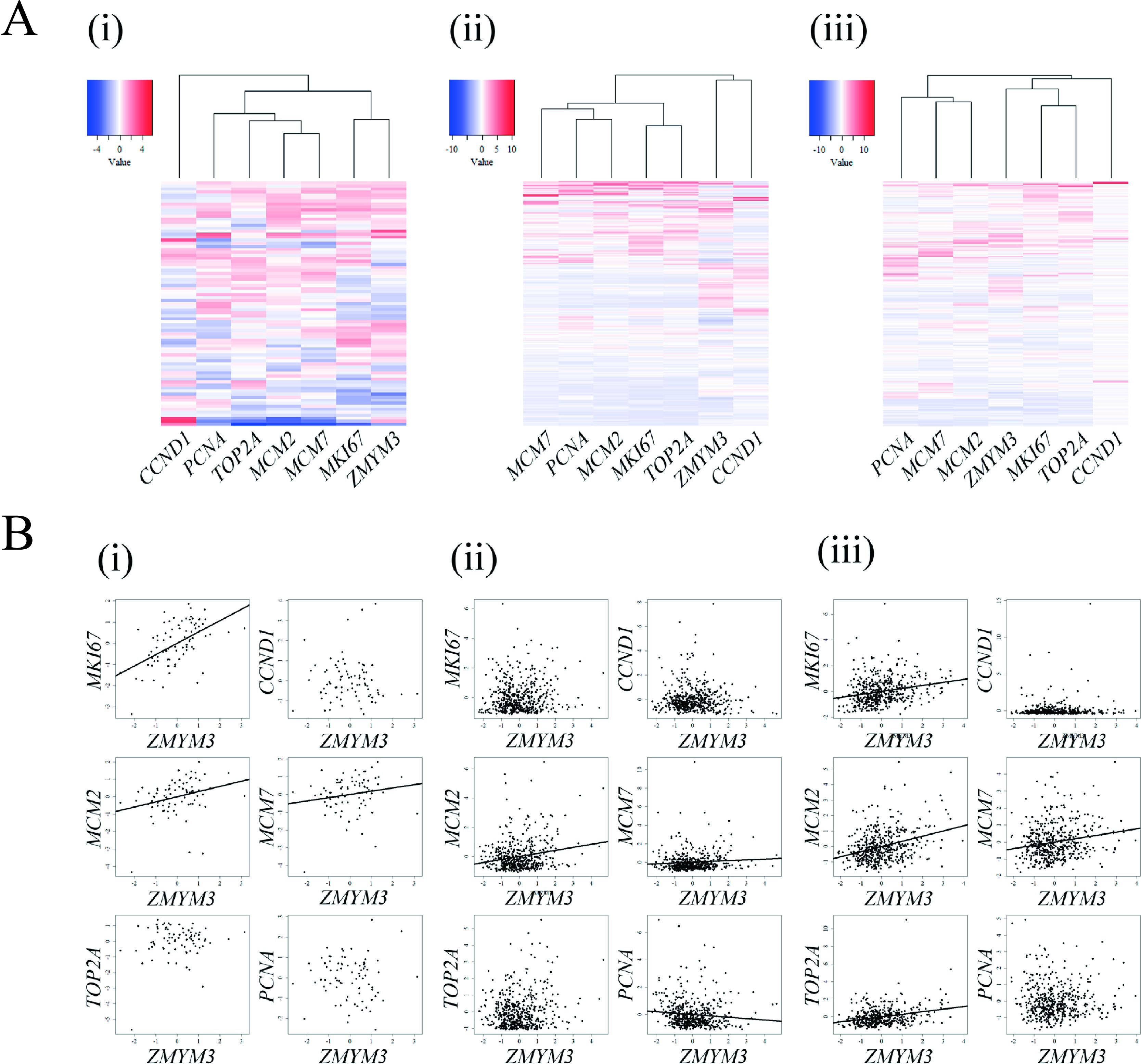
Zinc finger, myeloproliferative, and mental retardation-type containing 3 (ZMYM3) is a highly conserved protein among vertebrates. Although it promotes DNA repair and moderate histone acetylation, the other functions of ZMYM3 remain unclear. We herein examined the physiological functions of ZMYM3 in human lung cancer using a ZMYM3-knockdown small cell lung cancer (SCLC) cell line. ZMYM3-knockdown SCLC cells grew slowly and the Ki-67 labeling index was lower in ZMYM3-knockdown cells than in mock cells. The subcutaneous tumors that formed after xenotransplantation into immunodeficient mice were slightly smaller in the ZMYM3-knockdown group than in the mock group. Furthermore, public RNA-sequencing data analyses showed similar RNA profiles between ZMYM3 and some cell proliferation markers. These results indicate that ZMYM3 promotes cell proliferation in human lung carcinomas, particularly SCLC.

Keywords: ZMYM3; cell proliferation; immunohistochemistry; lung carcinoma; mouse tissues.


Conflict of interest statement

VThe authors declare that there are no conflicts of interest.

Figures

Fig. 1.
Fig. 1.
(A–D) ZMYM3 immunostaining in human lung tissues and lung cancers. (A) In normal tissues around tumors, bronchiolar epithelial cells (asterisk) and some alveolar cells (dagger), which appeared to be type II, were stained in nuclei. (B–D) In lung carcinomas, all 3 histological types: adenocarcinoma (ADC) (B), squamous cell carcinoma (SQC) (C), and small cell lung carcinoma (SCLC) (D), were strongly stained in nuclei. Original magnification: × 400. Bar = 50 μm. (E) Immunoblotting was also performed on 13 lung cancer cell lines: 7 SCLC cell lines (H69, H889, SBC-1, H69AR, H1688, SBC-3, and SBC-5) and 6 non-SCLC (NSCLC) cell lines (A549, H358, H1975, H226, H2170, and H15). All cell lines, except for H15, expressed ZMYM3. The prefix “NCI-” was omitted. β-actin was used as an internal control.
Fig. 2.
Fig. 2.
(A) The knockdown of ZMYM3 in the small cell lung carcinoma (SCLC) cell line, SBC-3. An immunoblotting analysis confirmed the decreased expression of ZMYM3. Daggers represent the cell clones used in the present study. β-actin was used as a loading control. (B) Immunoblotting of ZMYM3 knockdown cells and the negative control, mock cells, with a focus on cell proliferation. Cyclin D1 expression was slightly decreased in ZMYM3-knockdown cells. (C) Immunoblotting of ZMYM3-knockdown cells and mock cells with a focus on apoptosis. Under the induction of apoptosis by etoposide, cleaved caspase 3 increased in ZMYM3-knockdown cells. (D) Cell growth curve of the ZMYM3-knockdown cell clone and the mock. On day five, the cell count was significantly lower for the ZMYM3-knockdown cell clone than for the mock (average 465.0 versus 1153.0, p = 4.147 × 10⁻⁷). (E) Immunohistochemistry for ZMYM3 and Ki-67 in the ZMYM3-knockdown cell clone and the mock. Immunohistochemistry for ZMYM3 also confirmed the decreased expression of ZMYM3. The Ki-67 labeling index was significantly lower in the ZMYM3-knockdown cell clone than in the mock (average 416.4 versus 432.6 per 500 cells, p = 0.042). (F) The BrdU cell proliferation assay on the ZMYM3-knockdown cell clone and the mock. No significant differences were observed between the ZMYM3-knockdown cell clone and the mock. (G) Analysis of apoptosis by flow cytometry using annexin V/propidium iodide. Annexin V single-positive cells represent early apoptosis cells, while annexin V/propidium iodide double-positive cells represent late apoptosis cells. Even under the induction of apoptosis by etoposide, no marked differences were observed in apoptotic activities between ZMYM3-knockdown cell clone and the mock. KD: knockdown, CASP3: caspase 3.
Fig. 3.
Fig. 3.
Xenotransplantation of ZMYM3-knockdown cells and mock cells into the back skin of immunodeficient mice. (A) Tumors formed under the skin of mice. (B) The tumors of ZMYM3-knockdown cells were slightly smaller than those of mock cells. However, no significant differences were observed in volumes between ZMYM3-knockdown tumors and mock tumors (average 1181.5 versus 362.5, p = 0.222). (C) Hematoxylin-eosin staining of tumors. No significant morphological differences were observed between ZMYM3-knockdown tumors and mock tumors. Original magnification: × 400. Bar = 50 μm. (D) ZMYM3 and Ki-67 immunostaining in tumors. ZMYM3 immunostaining revealed a decrease in ZMYM3 expression in ZMYM3-knockdown cells. The Ki-67 labeling index was significantly lower in ZMYM3-knockdown cells than in mock cells (average 378.6 versus 350.8, p = 0.025). Original magnification: × 400. Bar = 50 μm. (E) TUNEL assay of tumors. No significant differences were noted in apoptosis counts between ZMYM3-knockdown tumors and mock tumors (average 6.6 versus 9.0, p = 0.373). Original magnification: × 200. Bar = 100 μm.
Fig. 4.
Fig. 4.
(A) Heat maps of RNA profiles in three histological subtypes of lung carcinoma: small cell lung carcinoma (SCLC) (i), adenocarcinoma (ADC) (ii), squamous cell carcinoma (SQC) (iii). (i, iii) In the heat maps of SCLC and SQC, the expression profiles of ZMYM3 were similar to those of cell proliferation markers (MKI67, MCM2, MCM7, TOP2A, and PCNA), but not to those of the cell cycle regulatory protein, CCND1. (ii) In contrast to SCLC and SQC, in the heat map of ADC, the similarity of expression profiles was weak between ZMYM3 and cell proliferation markers. (B) Scatter diagrams of ZMYM3 versus cell proliferation markers (MKI67, CCND1, MCM2, MCM7, TOP2A, and PCNA) in the three histological subtypes of lung carcinoma: SCLC (i), ADC (ii), and SQC (iii). From left to right, top to bottom: ZMYM3 versus MKI67, CCND1, MCM2, MCM7, TOP2A, and PCNA. When correlations were observed, regression lines were added to scatter diagrams. (i) Positive correlations were observed between ZMYM3 and MKI67 (ρ = 0.534, p = 2.894 × 10⁻⁷), MCM2 (ρ = 0.346, p = 1.640 × 10⁻³), and MCM7 (ρ = 0.227, p = 0.042). (ii) Positive correlations were observed between ZMYM3 and MCM2 (ρ = 0.098, p = 0.026) and MCM7 (ρ = 0.099, p = 0.025). A negative correlation was observed between ZMYM3 and PCNA (ρ = −0.150, p = 5.983 × 10⁻⁴). (iii) Positive correlations were observed between ZMYM3 and MKI67 (ρ = 0.264, p = 1.912 × 10⁻⁹), MCM2 (ρ = 0.264, p = 2.090 × 10⁻⁹), MCM7 (ρ = 0.176, p = 7.385 × 10⁻⁵), and TOP2A (ρ = 0.245, p = 2.773 × 10⁻⁸).

Similar articles

References

    1. Abeshouse, A., Ahn, J., Akbani, R., Ally, A., Amin, S., Andry, A. D., et al. (2015) The molecular taxonomy of primary prostate cancer. Cell 163; 1011–1025. - PMC - PubMed
    1. Alexander, M., Kim, S. Y. and Cheng, H. (2020) Update 2020: management of non‑small cell lung cancer. Lung 198; 897–907. - PMC - PubMed
    1. Alizadeh, F., Bozorgmehr, A., Tavakkoly-Bazzaz, J. and Ohadi, M. (2018) Skewing of the genetic architecture at the ZMYM3 human-specific 5' UTR short tandem repeat in schizophrenia. Mol. Genet. Genomics 293; 747–752. - PubMed
    1. Alizadeh, F., Moharrami, T., Mousavi, N., Yazarlou, F., Bozorgmehr, A., Shahsavand, E., et al. (2019) Disease-only alleles at the extreme ends of the human ZMYM3 exceptionally long 5' UTR short tandem repeat in bipolar disorder: a pilot study. J. Affect. Disord. 251; 86–90. - PubMed
    1. Aubry, S., Shin, W., Crary, J. F., Lefort, R., Qureshi, Y. H., Lefebvre, C., et al. (2015) Assembly and interrogation of Alzheimer’s disease genetic networks reveal novel regulators of progression. PLoS One 10; e0120352. - PMC - PubMed