Review
. 2021 Jul-Sep;15(3):283-299.
doi: 10.4103/sja.SJA_350_20. Epub 2021 Jun 19.

Anesthesia for thoracic surgery in infants and children

Affiliations
Review

Anesthesia for thoracic surgery in infants and children

Teresa M Murray-Torres et al. Saudi J Anaesth. 2021 Jul-Sep.

Abstract

The management of infants and children presenting for thoracic surgery poses a variety of challenges for anesthesiologists. A thorough understanding of the implications of developmental changes in cardiopulmonary anatomy and physiology, associated comorbid conditions, and the proposed surgical intervention is essential in order to provide safe and effective clinical care. This narrative review discusses the perioperative anesthetic management of pediatric patients undergoing noncardiac thoracic surgery, beginning with the preoperative assessment. The considerations for the implementation and management of one-lung ventilation (OLV) will be reviewed, and as will the anesthetic implications of different surgical procedures including bronchoscopy, mediastinoscopy, thoracotomy, and thoracoscopy. We will also discuss pediatric-specific disease processes presenting in neonates, infants, and children, with an emphasis on those with unique impact on anesthetic management.

Keywords: One-lung ventilation; pediatric anesthesia; thoracic surgery; thoracoscopy; thoracotomy.


Conflict of interest statement

There are no conflicts of interest.

Figures
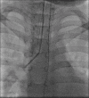
Figure 1
Figure 1
Fluoroscopic image showing the placement of a bronchial blocker in the right main stem bronchus. The light bend at the end of the blocker assists with guiding it into the desired bronchus
Figure 2
Figure 2
Fogarty® embolectomy catheter for use as a bronchial blocker during one-lung ventilation. These catheters have an inflatable, high pressure balloon at the tip that can be placed into and occlude the bronchus of the operative lung. They are available in a variety of sizes for use in a wide range of patient ages
Figure 3
Figure 3
Arndt® endobronchial blocker for one-lung ventilation. The Arndt® bronchial blocker is available in three sizes (5, 7, and 9 French) and features a central channel through which a wire is looped, to facilitate placement by a fiberoptic bronchoscope. When the wire loop is removed, the central channel can be used for suctioning or the administration of oxygen
Figure 4
Figure 4
Arndt Multi-port Adaptor® for introducing and securing the Arndt® blocker. The adaptor has four ports for: (1) attachment to the anesthesia circuit; (2) passage of the fiberoptic bronchoscope; (3) attachment to the endotracheal tube; and (4) entry of the bronchial blocker. The blocker port is tightened to secure the blocker in its final position
Figure 5
Figure 5
The Rusch® EZ blocker™ endobronchial blocker for one-lung ventilation. The Y-shaped 7 French catheter has a dual lumen system with two high-volume, low-pressure cuffs. It is positioned so that there is a balloon situated in each mainstem bronchus
Figure 6
Figure 6
Standard double-lumen endotracheal tube for one-lung ventilation. Double lumen endotracheal tubes (DLTs) are available in a variety of sizes, the smallest of which (26 French) may be suitable for patients as young as 8 years of age and 30 kg or greater in weight. The small internal lumens of the 26 French and 28 French DLTs can only accommodate ultrathin pediatric bronchoscopes
Figure 7
Figure 7
Adaptor for a double-lumen endotracheal tube. The adaptor has a standard 15 mm connector (1), and 2 ports (2) for passage of a fiberoptic bronchoscope or a suction catheter. The short segment of the tubing (black arrow) can be clamped during one-lung ventilation, and the port (2) is opened to allow the lung to deflate
Figure 8
Figure 8
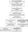
Flowchart for suggested management of oxygenation during one-lung ventilation. SpO2: Arterial oxygenation saturation by pulse oximetry; PaO2: Partial pressure of oxygen
Figure 9
Figure 9
Tracheoesophageal fistulae (arrows). There can be significant variation in the position, size, and shape of the opening of the fistula. Here, one fistula is located above the carina (left image) and one is located directly at the level of the carina (right image)
Figure 10
Figure 10
Anterior mediastinal mass. Computed tomography imaging demonstrates a large anterior mediastinal mass and severe tracheal compression (red arrow)

References

    1. Meneghini L, Zadra N, Zanette G, Baiocchi M, Giusti F. The usefulness of routine preoperative laboratory tests for one-day surgery in healthy children. Pediatr Anesth. 1998;8:11–5. - PubMed
    1. Nieto RM, De León LE, Diaz DT, Krauklis KA, Fraser CD., Jr Routine preoperative laboratory testing in elective pediatric cardiothoracic surgery is largely unnecessary. J Thorac Cardiovasc Surg. 2017;153:678–85. - PubMed
    1. Lee EY, Tracy DA, Mahmood SA, Weldon CB, Zurakowski D, Boiselle PM. Preoperative MDCT evaluation of congenital lung anomalies in children: Comparison of axial, multiplanar, and 3D images. AJR Am J Roentgenol. 2011;196:1040–6. - PubMed
    1. Baum VC, Barton DM, Gutgesell HP. Influence of congenital heart disease on mortality after noncardiac surgery in hospitalized children. Pediatrics. 2000;105:332–5. - PubMed
    1. Society BT. Guidelines on the selection of patients with lung cancer for surgery. Thorax. 2001;56:89–108. - PMC - PubMed