Review
. 2021 Sep 10;13(5):717-727.
doi: 10.1007/s12551-021-00841-6. eCollection 2021 Oct.

Current strategies of mechanical stimulation for maturation of cardiac microtissues

Affiliations
Review

Current strategies of mechanical stimulation for maturation of cardiac microtissues

Maria Carlos-Oliveira et al. Biophys Rev. .

Erratum in

Abstract

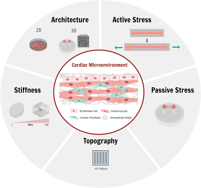
The most advanced in vitro cardiac models are today based on the use of induced pluripotent stem cells (iPSCs); however, the maturation of cardiomyocytes (CMs) has not yet been fully achieved. Therefore, there is a rising need to move towards models capable of promoting an adult-like cardiomyocytes phenotype. Many strategies have been applied such as co-culture of cardiomyocytes, with fibroblasts and endothelial cells, or conditioning them through biochemical factors and physical stimulations. Here, we focus on mechanical stimulation as it aims to mimic the different mechanical forces that heart receives during its development and the post-natal period. We describe the current strategies and the mechanical properties necessary to promote a positive response in cardiac tissues from different cell sources, distinguishing between passive stimulation, which includes stiffness, topography and static stress and active stimulation, encompassing cyclic strain, compression or perfusion. We also highlight how mechanical stimulation is applied in disease modelling.

Keywords: Cardiac microtissues; Maturation; Mechanical stimulation.


Figures

Fig. 1
Fig. 1
Cardiac microenvironment and mechanical stimuli applied aimed to mimic adult cardiac microtissue
Fig. 2
Fig. 2
Overview of different studies exploiting passive stimulation. A hESC-CMs were seeded onto PA substrates to study the effect of stiffness, cell anisotropy and cell-cell contact on cardiac maturation. (Rodriguez et al. 2019). B Microgrooves obtained through 3D printing were used to study the influence of surface topography on cell anisotropic re-arrangement. (Lind et al. 2017). C A scaffold-free approach with tri-culture (cardiomyocytes, fibroblasts and endothelial cells) was developed to study how passive stretch affects cardiac microtissue without the confounding effect of the biomaterial support. (Lui et al. 2021). D Progressive stretch (S3) over 3 weeks was compared to static stretch (S0) showing increased twitch force (Lu et al. 2021)
Fig. 3
Fig. 3
Overview of studies using active mechanical stimulation. A The so called mechano-active multielectrode-array, MaMEA allows the compression and stretch of cardiac microtissue integrated with electrodes (Imboden et al. 2019). B “BeatS-α” a 3D printed device capable of mechanical and electrical stimulation (Cortes et al. 2020). C Uniaxial stimulation was used to stimulate hiPSC-CMs (Kreutzer et al. 2020). D The beating-heart-on-chip platform cable to provide mechanical stimulus through a pneumatic platform (Marsano et al. 2016). E Cyclic compression was applied and compared with steady-state compression (Shachar et al. 2012)
Fig. 4
Fig. 4
Overview of different studies exploiting mechanical stimulation for disease modelling. A Rigid cantilevers (0.45 mN/mm) were seen to impair cardiomyocytes with titin mutation (Hinson et al. 2015). B Pressure and volume load were used to mimic hypertrophic and dilated cardiomyopathy (Rogers et al. 2019). C Cyclic stress was applied, and cardiac hypertrophy was shown as a result of the mechanical stimulus (Parsa et al. 2017). D 10% cyclic stress at 2–3Hz was used to mimic the failing myocardium (McCain et al. 2013)

References

    1. Bellin M, Mummery CL. Inherited heart disease – what can we expect from the second decade of human ips cell research? FEBS Lett. 2016;590:2482–2493. doi: 10.1002/1873-3468.12285. - DOI - PMC - PubMed
    1. Brandao KO, Tabel VA, Atsma DE, Mummery CL, Davis RP. Human pluripotent stem cell models of cardiac disease: from mechanisms to therapies. DMM Dis Models Mech. 2017;10(9):1039–1059. doi: 10.1242/dmm.030320. - DOI - PMC - PubMed
    1. Cashman TJ, Josowitz R, Johnson BV, Gelb BD, Costa KD. Human Engineered Cardiac Tissues Created Using Induced Pluripotent Stem Cells Reveal Functional Characteristics of BRAF-Mediated Hypertrophic Cardiomyopathy. PLoS ONE. 2016;11(1):1–17. doi: 10.1371/journal.pone.0146697. - DOI - PMC - PubMed
    1. Chang ACY, Pardon G, Chang ACH, Wu H, Ong SG, Eguchi A, Ancel S, Holbrook C, Ramunas J, Ribeiro AJS, LaGory EL, Wang H, Koleckar K, Giaccia A, Mack DL, Childers MK, Denning C, Day JW, Wu JC, Pruitt BL, Blau HM. Increased tissue stiffness triggers contractile dysfunction and telomere shortening in dystrophic cardiomyocytes. Stem Cell Rep. 2021;16:1–13. doi: 10.1016/j.stemcr.2021.04.018. - DOI - PMC - PubMed
    1. Cortes D, McTiernan CD, Ruel M, Franco W, Chu C, Liang W, Suuronen EJ, Alarcon EI (2020) BEaTS-α an open access 3D printed device for in vitro electromechanical stimulation of human induced pluripotent stem cells. Sci Rep 10(1). 10.1038/s41598-020-67169-1 - PMC - PubMed

LinkOut - more resources