

Case Reports
doi: 10.1016/j.radcr.2021.10.006.
eCollection 2022 Jan.
Diffuse axonal injury: a case report and MRI findings
Affiliations
- PMID: 34765068
- PMCID: PMC8571536
- DOI: 10.1016/j.radcr.2021.10.006
Item in Clipboard
Case Reports
Diffuse axonal injury: a case report and MRI findings
Radiol Case Rep.
.
Erratum in
-
Erratum regarding missing patient consent statements in previously published articles.Radiol Case Rep. 2023 Mar;18(3):1387-1388. doi: 10.1016/j.radcr.2022.10.050. Epub 2023 Jan 17. Radiol Case Rep. 2023. PMID: 36685799 Free PMC article.
Abstract
Diffuse axonal injury (DAI) is one of the most severe types of primary traumatic brain injury. In recent years, MR imaging has been gaining popularity as an adjunctive imaging method in patients with DAI. In this case report, we describe MRI findings of an 11-year-old male patient diagnosed with DAI and discuss the role of different sequences in the evaluation of DAI.
Keywords: Diffuse axonal injury; Diffusion tensor imaging; Diffusion weighted imaging; Magnetic resonance imaging; Susceptibility weighted imaging.
© 2021 The Authors. Published by Elsevier Inc. on behalf of University of Washington.
Figures
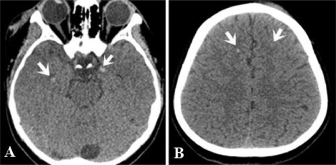
Multiple punctate hyperdense foci (microhemorrhage) at the gray-white matter junction of bilateral temporal (white arrow, A) and frontal lobes (white arrows, B)
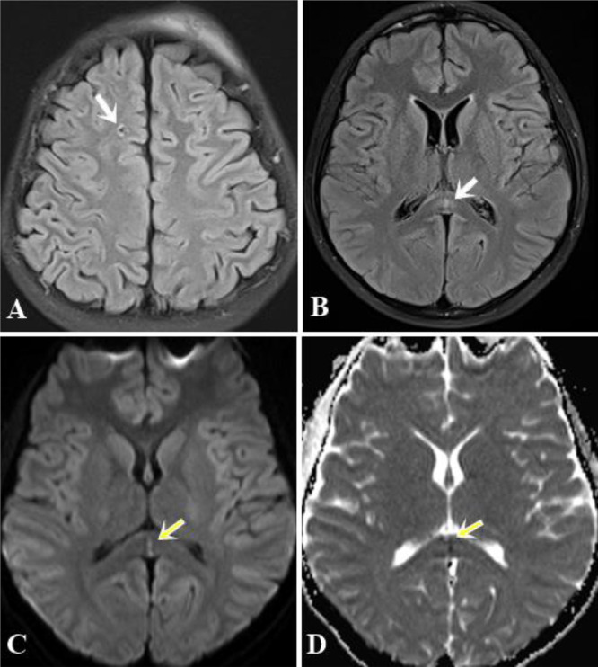
(A) and (B). Axial FLAIR images showed some punctate hyperintense foci in the subcortical white matter of the right frontal lobe (arrow) and the splenium of the corpus callosum (white arrows). (C) and (D). DWI and ADC maps indicated restricted diffusion within the splenium of the corpus callosum (yellow arrows) (Color version of figure is available online)
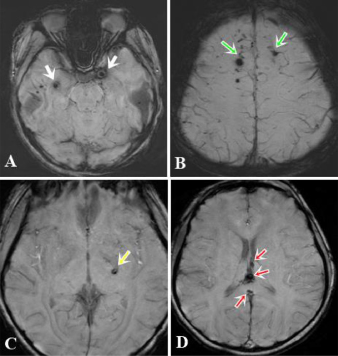
SWI illustrated multiple hypointense foci of hemorrhagic lesions at the grey-white matter junction of bilateral temporal lobes (white arrows, A), bilateral frontal lobes (green arrows, B), posterior limb of the left internal capsule (yellow arrow, C), the fornix commissure, and the splenium of the corpus callosum (red arrows, D) (Color version of figure is available online)
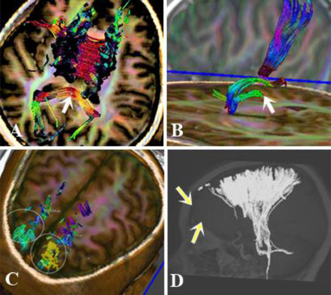
Diffusion Tensor Imaging with 3D-Fiber Tractography revealed disrupted white matter fibers in the posteroinferior aspect of the splenium (arrow, A), left crus of the fornix (arrow, B), and subcortical frontal tracts (circle C and arrow D)
References
-
- Adams JH, Graham DI, Murray LS, Scott G. Diffuse axonal injury due to nonmissile head injury in humans: an analysis of 45 cases. Ann neurol. 1982;12(6):557–563. - PubMed
-
- Adams JH, Doyle D, Ford I, Gennarelli TA, Graham DI, McLellan DR. Diffuse axonal injury in head injury: definition, diagnosis and grading. Histopathology. 1989;15(1):49–59. - PubMed
-
- Ashwal S, Babikian T, Gardner-Nichols J, Freier M-C, Tong KA, Holshouser BA. Susceptibility-weighted imaging and proton magnetic resonance spectroscopy in assessment of outcome after pediatric traumatic brain injury. Arch physic med rehabilitat. 2006;87(12):50–58. - PubMed
Publication types
LinkOut - more resources
Full Text Sources
