

doi: 10.1002/ajum.12241.
eCollection 2021 May.
Syncope and undifferentiated shock
Affiliations
- PMID: 34765417
- PMCID: PMC8412011
- DOI: 10.1002/ajum.12241
Item in Clipboard
Syncope and undifferentiated shock
Australas J Ultrasound Med.
.
Abstract
Ventricular free wall rupture is a rare post myocardial complication with a high associated mortality. In this article we discuss the case of an elderly patient who presented to our emergency department in shock after an episode of syncope. Using Point Of Care Ultrasound (POCUS), identification of cardiac tamponade and pericardial thrombus was possible, signs indicating a diagnosis of free wall rupture. Early initiation of transfer proceedings to a tertiary cardio-thoracic unit was therefore possible, resulting in a positive patient outcome.
© 2021 Australasian Society for Ultrasound in Medicine.
Conflict of interest statement
The authors have no commercial associations or sources of support that might pose a conflict of interest.
Figures
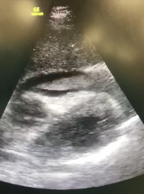
POCUS (RUSH protocol) demonstrating a large pericardial effusion, with a flaccid, hyperechogenic shadow within the pericardial cavity, left ventricular wall segmental akinesia and restricted expansion of the right ventricle. Additionally, paradoxical movement of the right atrium and dilation of the inferior vena cava were noted
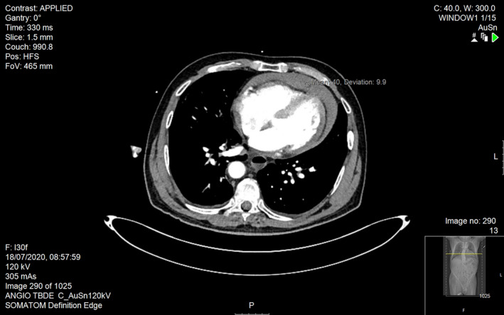
CTA revealed a medium pericardial effusion with thrombus formation, infarct within the left wall of the LV with mild pulmonary oedema and no evidence of aortic dissection
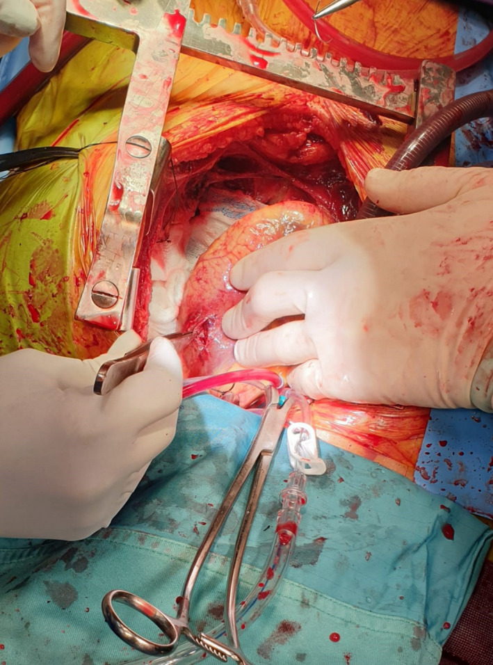
A photograph taken during the surgical procedure. After the clot was excavated from the pericardium, the tear was visualised and subsequently repaired. The size of the defect, as demonstrated at the operating theatre, was 3–4 cm
References
-
- Moreno R, López‐Sendón J, García E, de Isla Leopoldo P, de Sá Esteban L, Ortega A, et al. Primary angioplasty reduces the risk of left ventricular free wall rupture compared with thrombolysis in patients with acute myocardial infarction. J Am College Cardiol 2002; 39(4): 598–603. - PubMed
-
- Kutty RS, Jones N, Moorjani N. Mechanical complications of acute myocardial infarction. Cardiology Clin 2013; 31(4): 519–31. - PubMed
-
- Ibanez B, James S, Agewall S, Antunes MJ, Bucciarelli‐Ducci C, Bueno H, et al. 2017 ESC Guidelines for the management of acute myocardial infarction in patients presenting with ST‐segment elevation: The Task Force for the management of acute myocardial infarction in patients presenting with ST‐segment elevation of the European Society of Cardiology (ESC). ESC Scientific Document Group. Eur Heart J 2018; 39(2): 119–177. - PubMed
-
- McMullan MH, Maples MD, Kilgore TL, Hindman SH. Surgical experience with left ventricular free wall rupture. Ann Thoracic Surg 2001; 71(6): 1894–99. - PubMed
LinkOut - more resources
Full Text Sources