. 2021 Apr 7;11(6):481-492.
doi: 10.1016/j.jtcme.2021.03.005. eCollection 2021 Nov.

Inhibitory role of a smart nano-trifattyglyceride of Moringa oleifera root in epithelial ovarian cancer, through attenuation of FSHR - c-Myc axis

Affiliations

Inhibitory role of a smart nano-trifattyglyceride of Moringa oleifera root in epithelial ovarian cancer, through attenuation of FSHR - c-Myc axis

Arijit Ghosh et al. J Tradit Complement Med. .

Abstract

Background and aim: Epithelial ovarian cancer has the deadliest prognosis amongst gynaecological cancers, warranting an unmet need for newer drug targets. Based on its anticancer as well as abortifacient potential, Moringa oleifera Lam. root was hypothesized to have some implications in follicle stimulating hormone receptor (FSHR) dependent cancers like epithelial ovarian cancer.

Experimental procedure: Effect of Moringa oleifera Lam. root extract (MRE) was studied in epithelial ovarian cancer cell line through in vitro studies viz. MTT assay, clonogenic assay, cell cycle analysis, flow cytometry, western blot analysis, immunocytochemical analysis of FSHRand c-Myc expression and in vivo studies viz. effect of MRE in mice model of ovarian carcinoma. The structure of the active compound of MRE was elucidated following solvent extraction, purification through column chromatography, preparative TLC and bioactivity guided structural identification through 1H-NMR, 13C-NMR, DEPT-135, ESIMS,FT-IR spectrophotometry, UV-vis-NIR spectrophotometry and DFT study.

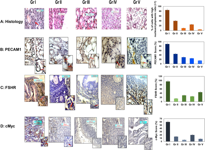
Results and conclusion: Crude MRE displayed cytotoxic activity, induced apoptosis, and attenuated expression of FSHR and c-Myc in ovarian cancer cell line OAW42. MRE also attenuated expression of CD31, FSHR, and c-Myc in tumour xenograft mouse model. Finally, the active compound purified from ethyl acetate-n-hexane subfraction ofMRE, that attenuated viability of ovarian carcinoma cell lines and reduced FSHR and c-Myc expression, was identified as a naturally hydrated-trifattyglyceride, showing aDFT-optimized folded amphipathic structure for easy transportation through hydrophilic and hydrophobic regions in a biological system, indicating its immense therapeutic relevance in epithelial ovarian carcinoma.

Keywords: Antitumor activity; Bioactivity guided structural identification; DEPT, Distortionless enhancement by polarization transfer; DFT, Density-functional theory; Density functional theory; Dynamic light scattering; ESI, Electrospray ionization; FSHR, follicle-stimulating hormone receptor; FT-IR, Fourier Transform Infrared Spectroscopy; MRE, Moringa root extract; Mice xenograft model; NMR, Nuclear Magnetic Resonance; Nuclear magnetic resonance; Scanning electron microscopy; UV-VIS-NIR, Ultraviolet–visible–near infrared spectroscopy.


Conflict of interest statement

The authors declare that they have no known competing financial interests or personal relationships that could have appeared to influence the work reported in this paper. The authors declare that they have no known competing financial interests or personal relationships that could have appeared to influence the work reported in this paper.

Figures

Image 1
Graphical abstract
Fig. 1
Fig. 1
In vitro study of Moringa oleifera Lam. root crude water extract (MRE) on survival of epithelial ovarian cancer cell line OAW42. Clonogenic assay (A, B) showed significant reduction of OAW42 colonies after treatment with 50% inhibitory concentration (IC-50: 250 μg/mL) of MRE, from data obtained from MTT cell viability assay (Fig. S1, S2). Cell cycle analysis showed a progressive increment of Propidium Iodide (PI) uptake in the subG0 phase from 24 h treatment, compared to the control (C, D). Statistical significance in PI uptake is being shown as bar diagram (E). Western Blot analysis for Apoptosis assay showing significant upregulation of cleaved caspase 9, 3 and PARP1, but not Caspase8, indicating MRE induced cell death of OAW42 cells through intrinsic apoptosis mechanism (F, G, H, I and J). The normalized fold change expression of candidate proteins showing MRE alone or in combination with Paclitaxel or Cisplatin, activated Caspase 9, Caspase 3, PARP1 expression, but not Caspase 8 (F–J). One-way ANOVA for statistical difference in survival of different treatment groups is shown in Table S1, S2 (p 0.05).
Fig. 2
Fig. 2
Immunocytochemical (A) and RNA expression (B) analyses of FSHR and c-Myc in OAW42 cells after treatment with MRE. Data showing expression, cellular localization and Mean Fluorescence intensities (C) of FSHR and c-Myc in OAW42 cells, upon treatment of MRE (250 μg/mL for 24 h; Magnifications 60X; Scale bar 10 μm. Densitometric fold change of expression normalized to GAPDH expression displaying significant reduction of expression of FSHR and c-Myc in MRE treated groups compared to control (D); densitometric analysis carried out using Biorad image lab software; U: untreated and T: treated for semiquantitative RT-PCR, a fold change ≤1.2 considering significant. One-way ANOVA for statistical difference in survival of different treatment groups is shown Table S3 (p ≤ 0.05).∗indicating statistical significance (p ≤ 0.05).
Fig. 3
Fig. 3
In vivo study of Moringa oleifera Lam. root crude water extract in xenograft mice model. Haematoxylene-Eosin stained mice ovarian tissue after day 30 (A; Fig. S3), showing different treatment groups, viz. Gr I: ovarian carcinoma control [OAW42+Cyclosporin A], Gr II: carcinoma group with MRE treatment [OAW42 + Cyclosporin A + MRE treated], Gr III: only MRE treated, Gr IV: only Cyclosporin A treated, Gr V: untreated. For ovarian carcinoma control group, high nucleocytoplasmic index (black arrow) evidenced (A: Gr I); magnification 40X; scale bar: 50 μM. Immunohistochemical localization of CD31 (PECAM1) (B), FSHR (C) and c-Myc (D) in ovarian cortex/ampulla region in ovarian carcinoma control group (Gr I) was compared with other four groups (n = 3 each). (Magnification 20X; inset 40X; scalebar100 μM). Arrow indicates expression. A significantly increased propensity of cells with high nucleocytoplasmic index, high expression of PECAM1, FSHR and c-Myc was evident in Gr I mice, compared to MRE treated and control groups. One-way ANOVA for statistical difference in survival of different treatment groups is shown Table S4 (p 0.05).∗.
Fig. 4
Fig. 4
Immunocytochemical and RNA expression analysis of FSHR and cMyc in OAW42 and OVCAR3 cells after treatment with purified active subfraction of MRE: Immunocytochemical analysis of FSHR and cMyc in (A) OAW42 and (B) OVCAR3 cells and their mean fluorescence intensities (C) and (D) respectively, with/without treatment with purified active 10% ethyl acetate-n hexane subfraction of MRE (designated as TG). Magnifications 60X. Scale bar represent 10 μm. Arrow heads indicate expression. Mean fluorescence intensity calculation showed significant reduction of expression of FSHR and c-Myc in treated cells (C, D). In treatment groups, intensity of FSHR and c-Myc was reduced, diffusely cytoplasmic and non-nucleolar. One-way ANOVA for statistical difference in survival of different treatment groups is shown in supplementary Table S5 (p 0.05). ∗ Indicate statistical significance. RNA expression analysis of FSHR and c-Myc proteins in OAW42 and in OVCAR3 cells respectively with/without treatment with active fraction F4 of MRE by semiquantitative RT-PCR method (E, F). The band intensities of each gene were normalized to GAPDH gene in both cell lines, using Biorad Image lab software (E, F). For semiquantitative RT-PCR in OAW42 and OVCAR3 respectively, a fold change of ≤ 1.2 was considered significant. ∗ indicate statistical significance (p 0.05) (G, H, Supplementary Table S5B).
Fig. 5
Fig. 5
Characterization, nano-scale property, transportation, and DFT study of hydrated-triglyceride. (A) 1H NMR spectrum of the drug with chemically two non-equivalent protons. (B) ESI-MS spectrum of water bound drug. (C) UV–vis and (D) DLS spectrum. (E) SEM image of the as-extracted drug after sample preparation by spin coating method. Optimized geometrical morphology of the drug in water (F) and cyclohexane (G). (H) Model for easy transportation of the hydrated-triglyceride through the aqueous exterior, interior and lipophilic cell membrane.

References

    1. Webb P.M., Jordan S.J. Epidemiology of epithelial ovarian cancer. Best Pract Res Clin Obstet Gynaecol. 2017;41:3–14. doi: 10.1016/j.bpobgyn.2016.08.006. - DOI - PubMed
    1. Koshiyama M., Matsumura N., Konishi I. Recent concepts of ovarian carcinogenesis: type I and type II. BioMed Res Int. 2014;934261:1–11. doi: 10.1155/2014/934261. - DOI - PMC - PubMed
    1. Bose C.K. Anomalous endocrine feedback of peri-menopause in the etiology of type II ovarian cancer. Future Oncol. 2013;9:1257–1261. doi: 10.2217/fon.13.147. - DOI - PubMed
    1. Choi J.H., Choi K.C., Auersperg N., Leung P.C. Overexpression of follicle-stimulating hormone receptor activates oncogenic pathways in preneoplastic ovarian surface epithelial cells. J Clin Endocrinol Metab. 2004;89:5508–5516. doi: 10.1210/jc.2004-0044. - DOI - PubMed
    1. Omara T., Kiprop A.K., Ramkat R.C., et al. Medicinal plants used in traditional management of cancer in Uganda: a review of ethnobotanical surveys, phytochemistry, and anticancer studies. Evidence-Based Complementary and Alternative Medicine. Evid Based Complementary Altern Med. 2020;2020 doi: 10.1155/2020/3529081. - DOI - PMC - PubMed

LinkOut - more resources