. 2021 Oct 26:8:744779.
doi: 10.3389/fcvm.2021.744779. eCollection 2021.

Late Gadolinium Enhancement Cardiovascular Magnetic Resonance Assessment of Substrate for Ventricular Tachycardia With Hemodynamic Compromise

Affiliations

Late Gadolinium Enhancement Cardiovascular Magnetic Resonance Assessment of Substrate for Ventricular Tachycardia With Hemodynamic Compromise

John Whitaker et al. Front Cardiovasc Med. .

Abstract

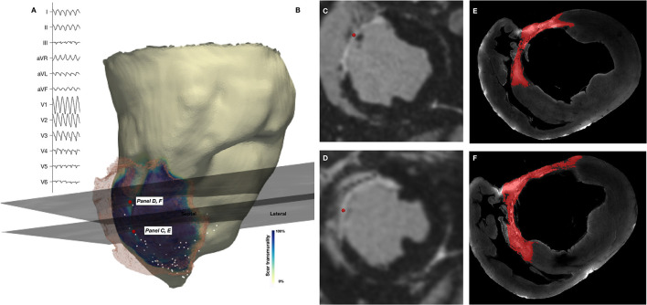
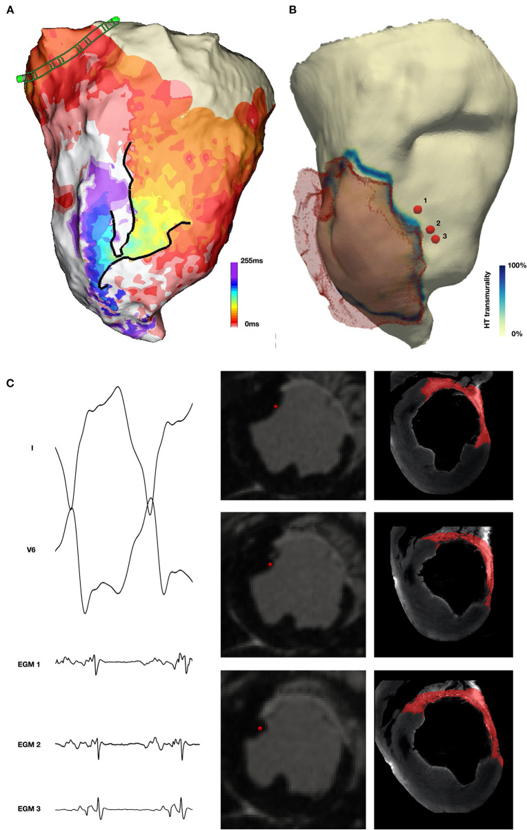
Background: The majority of data regarding tissue substrate for post myocardial infarction (MI) VT has been collected during hemodynamically tolerated VT, which may be distinct from the substrate responsible for VT with hemodynamic compromise (VT-HC). This study aimed to characterize tissue at diastolic locations of VT-HC in a porcine model. Methods: Late Gadolinium Enhancement (LGE) cardiovascular magnetic resonance (CMR) imaging was performed in eight pigs with healed antero-septal infarcts. Seven pigs underwent electrophysiology study with venous arterial-extra corporeal membrane oxygenation (VA-ECMO) support. Tissue thickness, scar and heterogeneous tissue (HT) transmurality were calculated at the location of the diastolic electrograms of mapped VT-HC. Results: Diastolic locations had median scar transmurality of 33.1% and a median HT transmurality 7.6%. Diastolic activation was found within areas of non-transmural scar in 80.1% of cases. Tissue activated during the diastolic component of VT circuits was thinner than healthy tissue (median thickness: 5.5 mm vs. 8.2 mm healthy tissue, p < 0.0001) and closer to HT (median distance diastolic tissue: 2.8 mm vs. 11.4 mm healthy tissue, p < 0.0001). Non-scarred regions with diastolic activation were closer to steep gradients in thickness than non-scarred locations with normal EGMs (diastolic locations distance = 1.19 mm vs. 9.67 mm for non-diastolic locations, p < 0.0001). Sites activated late in diastole were closest to steep gradients in tissue thickness. Conclusions: Non-transmural scar, mildly decreased tissue thickness, and steep gradients in tissue thickness represent the structural characteristics of the diastolic component of reentrant circuits in VT-HC in this porcine model and could form the basis for imaging criteria to define ablation targets in future trials.

Keywords: cardiovascular magnetic resonance; late gadolinium enhancement; mechanical circulatory support; venous-arterial extra corporeal membrane oxygenation (VA-ECMO); ventricular tachycardia.


Conflict of interest statement

The CardioHelp machine and all ECMO consumables were donated by Maquet, Getinge Group without conditions on their use. The Precision and Claris system and all EP mapping consumables were donated by Abbott without conditions on their use. The authors declare a potential conflict of interest and state it below. RN was employed by the company Siemens Healthcare. SK and TA were employed by the company Abbott Medical. The remaining authors declare that the research was conducted in the absence of any commercial or financial relationships that could be construed as a potential conflict of interest.

Figures

Figure 1
Figure 1
(A) Activation map during right ventricular (RV) pacing at 500 ms. Yellow dots mark the point at which activation time was recorded. Black line represents linear conduction block during pacing at this cycle length. (B) Activation map during right ventricular (RV) pacing at 300 ms. (C) Number of distinct regions of conduction block during different heart rhythms (RV pacing at 500 ms and 300 ms and during VT). (D) Scatter plot of extent of linear endocardial conduction block and activation rate during pacing or VT.
Figure 2
Figure 2
Visualization of location of diastolic EGMs on in-vivo CMR. Column (A) and column (B) show short axis (SAX) and long axis (LAX) multiplanar reconstruction (MPR) of in-vivo CMR with the location of the corresponding diastolic EGM [column (D)] indicated by a red sphere. Column (C) shows SAX MPR of in-vivo CMR with segmentation of scar (yellow) and heterogeneous tissue (HT) (red) superimposed.
Figure 3
Figure 3
Co-localization of diastolic locations from Electroanatomic Mapping System and in-vivo imaging and episcopic auto-fluorescence cryomicrotome imaging. (A) Surface 12-lead ECG of induced VT. (B) In-vivo CMR derived shell color coded according to scar transmurality, with translucent mesh derived from scar also shown. Location of 2 diastolic EGMs is shown (red spheres) and corresponding short axis (SAX) in-vivo LGE CMR slice. (C,D) SAX slices shown in (B), with location of recorded EGM (red sphere). (E,F) Corresponding EACI data with segmented scar (red) superimposed.
Figure 4
Figure 4
Localization of diastolic locations on in-vivo CMR imaging. (A) Activation map demonstrating re-entrant VT with a figure 8 pattern of re-entry. (B) CMR derived mesh color coded according to scar transmurality with location of selected diastolic EGMs indicated by red sphere and corresponding in-vivo LGE CMR imaging indicated. (C) Diastolic EGM and corresponding location on in-vivo CMR at four sequential points along the diastolic isthmus of this VT.
Figure 5
Figure 5
Tissue characteristics of diastolic locations. (A) Example of LGE CMR derived endocardial shell color coded according to scar transmurality and demonstrating antero-septal infarction. Histogram shows combined scar/heterogeneous tissue (HT) transmurality pooled from all locations activated during diastole in all pigs. (B) Example of LGE CMR derived endocardial shell color coded according to tissue thickness. Histogram shows tissue thickness pooled from all diastolic locations (blue) in all pigs compared with tissue thickness at the location of normal EGMs pooled from all pigs (red). (C) Example of LGE CMR derived endocardial shell color coded according to distance from HT. Histogram shows distance from HT pooled from all diastolic locations in six pigs.
Figure 6
Figure 6
Non-scarred tissue demonstrating diastolic activation during VT. (A) Activation map during VT demonstrating diastolic activation during VT (yellow to purple) with a converging pattern of activation toward a narrow channel (green) which subsequently widens prior to multiple systolic breakouts (red). (B) In-vivo CMR derived shell with translucent mesh of scar overlaid and color coded according to HT transmurality. The location of 3 early diastolic EGMs is indicated by red spheres. (C) Early diastolic EGM, short axis (SAX) in-vivo CMR and corresponding EACI imaging at locations 1, 2 and 3 demonstrating location of EGMs in tissue proximal to scar, but without enhancement in the wall at this location.

References

    1. Mehra R, Zeiler RH, Gough WB, El-Sherif N. Reentrant ventricular arrhythmias in the late myocardial infarction period. 9 electrophysiologic-anatomic correlation of reentrant circuits. Circulation. (1983) 67:11–24. 10.1161/01.CIR.67.1.11 - DOI - PubMed
    1. El-Sherif N, Smith RA, Evans K. Canine ventricular arrhythmias in the late myocardial infarction period. 8 epicardial mapping of reentrant circuits. Circ Res. (1981) 49:255–65. 10.1161/01.RES.49.1.255 - DOI - PubMed
    1. Qin D, Zhang Z-H, Caref EB, Boutjdir M, Jain P, El-Sherif N. Cellular and ionic basis of arrhythmias in postinfarction remodeled ventricular myocardium. Circ Res. (1996) 79:461–73. 10.1161/01.RES.79.3.461 - DOI - PubMed
    1. Stevenson WG, Friedman PL, Sager PT, Saxon LA, Kocovic D, Harada T, et al. . Exploring postinfarction reentrant ventricular tachycardia with entrainment mapping. J Am Coll Cardiol. (1997) 29:1180–9. 10.1016/S0735-1097(97)00065-X - DOI - PubMed
    1. Tung R, Mathuria N, Michowitz Y, Yu R, Buch E, Bradfield J, et al. . Functional pace-mapping responses for identification of targets for catheter ablation of scar-mediated ventricular tachycardia. Circ Arrhythmia Electrophysiol. (2012) 5:264–72. 10.1161/CIRCEP.111.967976 - DOI - PMC - PubMed

LinkOut - more resources