Review
. 2021 Sep 1;3(21):6007-6026.
doi: 10.1039/d1na00536g. eCollection 2021 Oct 27.

Emergence of cationic polyamine dendrimersomes: design, stimuli sensitivity and potential biomedical applications

Affiliations
Review

Emergence of cationic polyamine dendrimersomes: design, stimuli sensitivity and potential biomedical applications

Partha Laskar et al. Nanoscale Adv. .

Abstract

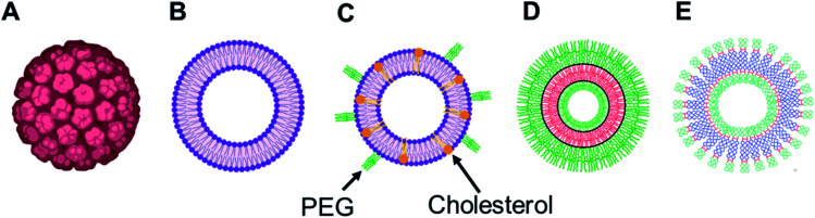
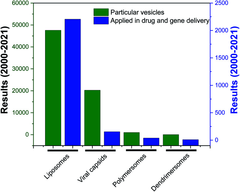
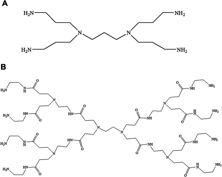
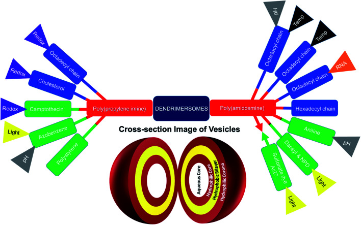
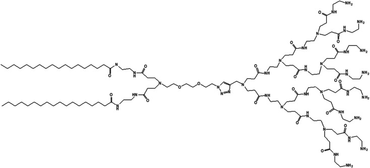
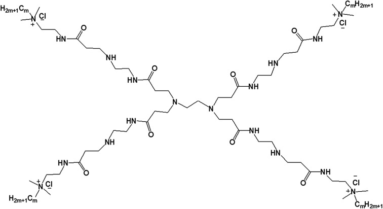
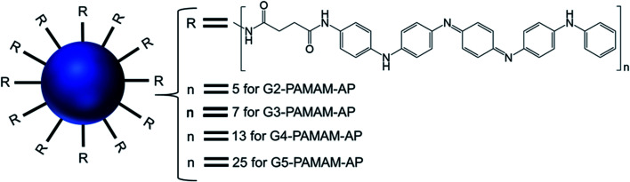
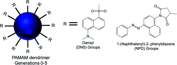
For decades, self-assembled lipid vesicles have been widely used in clinics as nanoscale delivery systems for various biomedical applications, including treatment of various diseases. Due to their core-shell architecture and versatile nature, they have been successfully used as carriers for the delivery of a wide range of therapeutic cargos, including drugs and nucleic acids, in cancer treatment. Recently, surface-modified polyamine dendrimer-based vesicles, or dendrimersomes, have emerged as promising alternatives to lipid vesicles for various biomedical applications, due to their ease of synthesis, non-immunogenicity, stability in circulation and lower size polydispersity. This mini-review provides an overview of the recent advances resulting from the use of biomimetic hydrophobically-modified polyamine-based dendrimersomes towards biomedical applications, focusing mainly on the two most widely used polyamine dendrimers, namely polyamidoamine (PAMAM) and poly(propylene imine) (PPI) dendrimers.


Conflict of interest statement

There are no conflicts to declare.

Figures

Fig. 1
Fig. 1. Representation of various vesicular structures: (A) viral capsid, (B) liposome (or lipid-based vesicle), (C) stealth liposome (with additives such as cholesterol and PEG), (D) polymersome (or polymer-based vesicle), (E) dendrimersome (or dendrimer-based vesicle). For clarity, only single-bilayer vesicles were represented in this figure.
Fig. 2
Fig. 2. Research articles cited in PubMed (search date: 10th March 2021) published from 2000 to 2021, corresponding to the keywords “liposomes”, “viral capsids”, “polymersomes”, “dendrimersomes” (for the category “particular vesicles”) and “liposomes in drug and gene delivery”, “viral capsids in drug and gene delivery”, “polymersomes in drug and gene delivery”, “dendrimersomes in drug and gene delivery” (for the category “application in drug and gene delivery”).
Fig. 3
Fig. 3. Chemical structure of generation 1-diaminobutyric polypropylenimine (PPI) (A) and generation 1-polyamidoamine (PAMAM) dendrimers (B).
Fig. 4
Fig. 4. Surface-modified amphiphilic dendrimers forming stimuli-responsive and non-stimuli-responsive dendrimer-based vesicles, or dendrimersomes, presented in this review (color code for surface-modifying groups: lipids or aliphatic chains (blue), aromatic groups (green); “double colored bar”: conjugated amphiphiles or hydrophobe–dendrimer conjugate, “→←”: complexed amphiphiles or hydrophobe–dendrimer complex).
Fig. 5
Fig. 5. Chemical structure of an amphiphilic, surface-modified, disulphide-linked octadecyl chain-bearing/cholesterol-bearing/camptothecin-bearing PEGylated generation 3-diaminobutyric polypropylenimine (PPI or DAB) dendrimer (DAB-PEG-S-S-ODT/DAB-PEG-S-S-CHOL/DAB-PEG-S-S-CPT respectively), where m = 45.
Fig. 6
Fig. 6. (A) Measurement of CAC from the plot of relative fluorescence intensity (I/I0) of a hydrophobic fluorescent dye, N-phenyl-1-naphthylamine in presence of various lipid–dendrimer concentration in PBS buffer (pH 7.4). (B) Stained TEM images of lipid–dendrimer based dendrimersomes (400 μg mL−1 in PBS, pH 7.4) (bar size: 100 nm). (C) Size (■) and zeta potential (○) of dendrimersomes at various lipid–dendrimer concentrations in PBS buffer (pH 7.4) at 37 °C. AFM peak force error and height sensor images of 5 × 5 μm surfaces of (D) 200 μg mL−1 and (E) 500 μg mL−1 dendrimersomes adsorbed on a mica (silicon) surface (reproduced from ref. , with permission from the journal as authors of this article).
Fig. 7
Fig. 7. Unstained TEM images of low-cholesterol (A) and high-cholesterol (B) dendrimersomes (400 μg mL−1 in PBS (pH 7.4)). Unstained TEM images of dendrimersomes–DNA complexes: low-cholesterol (C and D) and high-cholesterol (E and F) dendriplexes at dendrimer : DNA weight ratios of 5 : 1 (C and E) and 10 : 1 (D and F) (reproduced from ref. with permission from the journal as authors of this article).
Fig. 8
Fig. 8. TEM images of dendrimersomes formed by CPT–dendrimer solutions (pH 7.4) at various concentrations: 0.2 mg mL−1 (A), 1.0 mg mL−1 (B) and 2.0 mg mL−1 (C) (scale bar: 100 nm). (D) Confocal microscopy images of the cellular uptake of CPT–dendrimer conjugate complexed with Cy5-labelled DNA (2.5 μg per well) at various weight ratios (control : DAB dendrimer : DNA) (magnification: ×40) (reproduced from ref. with permission from the journal as authors of this article).
Fig. 9
Fig. 9. Chemical structure of the surface-modified dendritic amphiphiles made of generation 5-poly(propylenimine) dendrimers 1, 2, and 3, with the dendrimers decorated with side chains containing 64 palmitoyl, 32 palmitoyl, 32 azobenzene and 64 azobenzene groups, respectively.
Fig. 10
Fig. 10. Chemical structure of polystyrene conjugated to the highest generation poly(propylene imine) dendrimer PS-dendr-(NH2)32.
Fig. 11
Fig. 11. Chemical structure of amine-terminated, generation 1-polyamidoamine dendron bearing two octadecyl hydrophobic chains.
Fig. 12
Fig. 12. Chemical structure of (A) generation 2- and (B) generation 3-PAMAM dendrimer conjugated to two octadecyl chains (R = isobutyramide (IBAM) or acetamide (ACAM) terminal groups).
Fig. 13
Fig. 13. Chemical structure of lipid-modified amphiphilic PAMAM dendrimer (AD) obtained by conjugating two precursors (azide-functionalized hydrophilic dendrimer moiety and alkyne-functionalized C-18 alkyl bearing hydrophobic moiety) through click chemistry.
Fig. 14
Fig. 14. Chemical structure of amphiphilic lipid-modified PAMAM dendrimers GnQPAMCm (where n = 1 (dendrimer generation) and m = 8, 12, 16 (alkyl chain length after the modification of the end group)).
Fig. 15
Fig. 15. Structure of aniline pentamer (AP) conjugated to various generations (G2–5) of PAMAM dendrimer, forming PAMAM-AP amphiphiles (where R = aniline pentamer, n = average number of AP groups).
Fig. 16
Fig. 16. Chemical structure of modified generation 0 to 5 PAMAM dendrimers conjugated to hydrophobic aromatic dansyl (DNS) or 1-(naphthalenyl)-2-phenyldiazene (NPD) groups, via sulfonylation and a Michael addition reaction respectively.
Fig. 17
Fig. 17. Chemical structure of the cationic generation-8 PAMAM dendrimer and the anionic trivalent sulfonate dye Ar27.
None
Partha Laskar
None
Christine Dufès

References

    1. Dufès C. Uchegbu I. F. Schätzlein A. G. Adv. Drug Delivery Rev. 2005;57:2177–2202. doi: 10.1016/j.addr.2005.09.017. - DOI - PubMed
    1. Paleos C. M. Tsiourvas D. Sideratou Z. Mol. Pharm. 2007;4:169–188. doi: 10.1021/mp060076n. - DOI - PubMed
    1. Somani S. Dufès C. Nanomedicine. 2014;9:2403–2414. doi: 10.2217/nnm.14.130. - DOI - PubMed
    1. Wu L. P. Ficker M. Christensen J. B. Trohopoulos P. N. Moghimi S. M. Bioconjugate Chem. 2015;26:1198–1211. doi: 10.1021/acs.bioconjchem.5b00031. - DOI - PubMed
    1. Wang B. B. Zhang X. Jia X. R. Li Z. C. Ji Y. Yang L. Wei Y. J. Am. Chem. Soc. 2004;126:15180–15194. doi: 10.1021/ja048219r. - DOI - PubMed