Review
. 2021 Oct 14;2(4):100174.
doi: 10.1016/j.xinn.2021.100174. eCollection 2021 Nov 28.

Nanotechnology-enhanced immunotherapy for metastatic cancer

Affiliations
Review

Nanotechnology-enhanced immunotherapy for metastatic cancer

Peisen Zhang et al. Innovation (Camb). .

Abstract

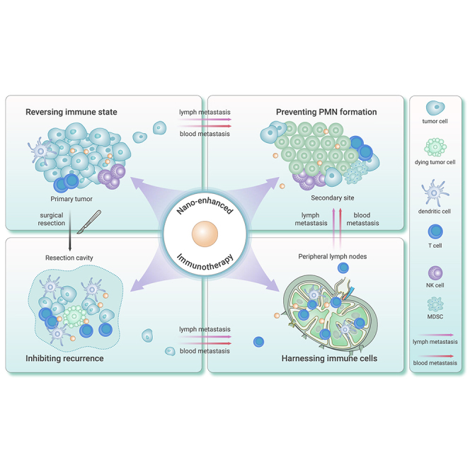
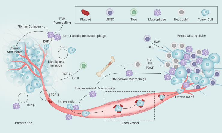
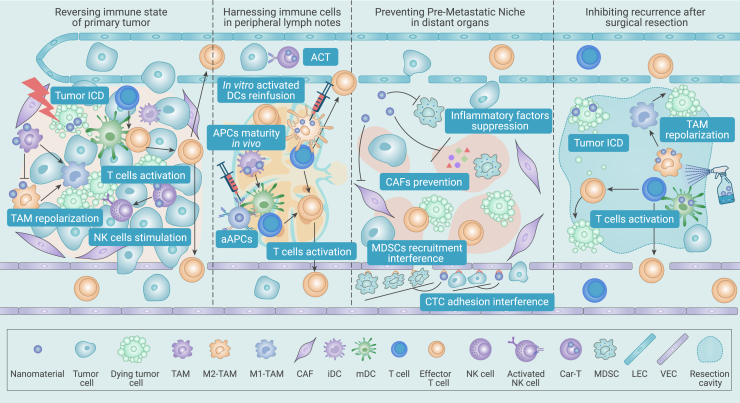
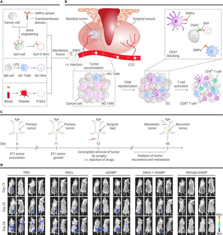
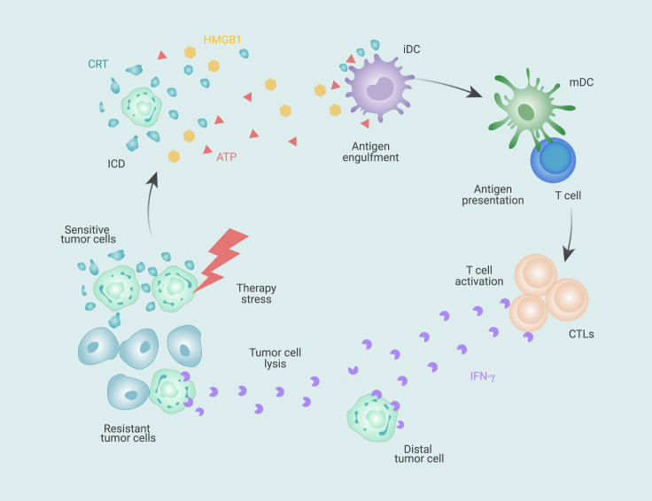
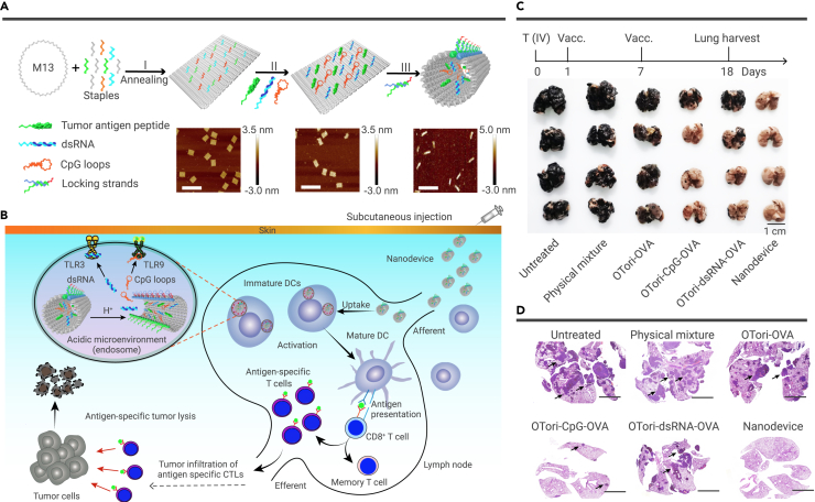
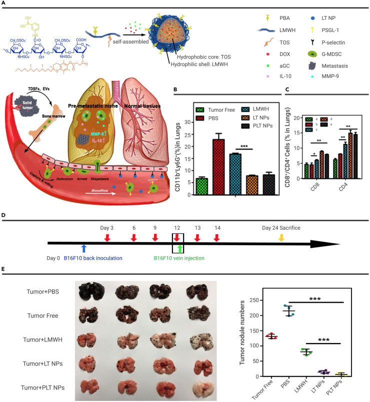
A vast majority of cancer deaths occur as a result of metastasis. Unfortunately, effective treatments for metastases are currently lacking due to the difficulty of selectively targeting these small, delocalized tumors distributed across a variety of organs. However, nanotechnology holds tremendous promise for improving immunotherapeutic outcomes in patients with metastatic cancer. In contrast to conventional cancer immunotherapies, rationally designed nanomaterials can trigger specific tumoricidal effects, thereby improving immune cell access to major sites of metastasis such as bone, lungs, and lymph nodes, optimizing antigen presentation, and inducing a persistent immune response. This paper reviews the cutting-edge trends in nano-immunoengineering for metastatic cancers with an emphasis on different nano-immunotherapeutic strategies. Specifically, it discusses directly reversing the immunological status of the primary tumor, harnessing the potential of peripheral immune cells, preventing the formation of a pre-metastatic niche, and inhibiting the tumor recurrence through postoperative immunotherapy. Finally, we describe the challenges facing the integration of nanoscale immunomodulators and provide a forward-looking perspective on the innovative nanotechnology-based tools that may ultimately prove effective at eradicating metastatic diseases.

Keywords: immunomodulators; immunotherapy; metastatic cancer; nanomaterials; tumor microenvironments.


Conflict of interest statement

The authors declare no competing interest.

Figures

None
Graphical abstract
Figure 1
Figure 1
The microenvironment promotes metastasis Host immune cells, together with various cytokines and growth factors secreted by tumor cells, immune cells, and stromal cells, support the dissemination and colonization of tumor cells in the pre-metastatic niche. Note: this figure is modified from Quail and Joyce.
Figure 2
Figure 2
Overview of this article Nano-enabled immunotherapy for inhibiting tumor metastasis and recurrence, including changing immune activity within the primary tumor, activating the peripheral immune system, interrupting the pre-metastasis niche, and conferring immunity to tumor cells to inhibit recurrence after surgery.
Figure 3
Figure 3
Hybrid cellular membrane nanovesicles amplify macrophage immune responses against cancer recurrence and metastasis (A) Schematic displaying hNVs consisting of engineered SαV-C-NVs, M1-NVs, and P-NVs. (B) Schematic displaying the interaction between hNVs and CTCs in the blood, accumulation in the postsurgical tumor bed, repolarization of TAMs toward an M1 phenotype, and blockage of the CD47-SIRPα “don’t eat me” pathway, which promotes the phagocytosis of cancer cells by macrophages and boosts antitumor T cell immunity. (C) Schematic displaying the treatment implemented in a mouse model of cancer recurrence after incomplete resection. (D) In vivo bioluminescence imaging of B16F10 tumor recurrence in different treatment groups. Reproduced with permission from Rao et al. Copyright 2020 Nature Publishing Group.
Figure 4
Figure 4
Tumor ICD As a result of endoplasmic reticulum (ER) stress and autophagy, dying cancer cells respond to therapeutic stress by exposing calreticulin (CRT) on their outer membrane at the pre-apoptotic stage and releasing ATP and the nuclear protein high-mobility group box 1 (HMGB1) during apoptosis. CRT, ATP, and HMGB1 bind to the receptor of immature dendritic cells (iDC), which facilitates the recruitment of iDCs into the tumor bed (stimulated by ATP), the engulfment of tumor antigens by iDCs (stimulated by CRT), and antigen presentation to T cells (stimulated by HMGB1). Activated CTLs will secret IFN-γ, which eventually leads to the inhibition of therapy-resistant tumor cells and distal tumor cells that therapeutics have not directly reached.
Figure 5
Figure 5
Nanoparticle-enhanced radiotherapy to trigger robust cancer immunotherapy for metastasis prevention (A) Antitumor immune response induced by PLGA-R837@Cat radiotherapy and checkpoint blockade to inhibit metastasis and recurrence. (B) Inhibition of tumor metastasis by radiotherapy with PLGA-R837@Cat plus αCTLA4 therapy in a 4T1 orthotopic breast tumor metastasis model. (C) Morbidity-free survival of different groups of mice with metastatic 4T1 tumors after various treatments. (∗ P < 0.05). (D) In vivo bioluminescence images showing the spreading and growth of firefly luciferase-4T1 (fLuc-4T1) cancer cells in different groups of mice after eliminating their primary orthotopic tumors. Reproduced with permission from Chen et al. Copyright 2019, Wiley-VCH.
Figure 6
Figure 6
Engineering magnetosomes for high-performance cancer vaccination (A) Fabrication process of A/M/C-MNC. (B) Schematic illustration of A/M/C-MNC-mediated cellular immune responses to elicit CTLs and memory T cells (TM cells) for cancer immunotherapy. (C) Bioluminescence images of hematogenous metastasis in lungs after intravenous inoculation of luciferase-expressing 4T1 (Luc-4T1) cells with different pretreatments: (I) PBS; (II) MF; (III) M-MNC; (IV) M/C-MNC; (V) A/M/C-MNC; (VI) A/M/C-MNC with magnetic retention; (VII) A/M/C-MNC with magnetic retention and anti-PD-1. (D) MFI statistics of lung metastasis in (C). (mean ± SD, n = 6, ∗∗∗P < 0.01) (E) Survival rate of mice in (C). Reproduced with permission from Li et al. Copyright 2019, American Chemical Society.
Figure 7
Figure 7
Nano-immunopotentiators Fe3O4-OVA promote cancer immunotherapy for preventing lung metastasis of melanoma (A) Schematic illustration of the Fe3O4-OVA vaccination strategy. The Fe3O4-OVA vaccine composed of ultra-small Fe3O4 nanoparticles and OVA not only improved DC maturation and T cell activation but also showed a positive effect on macrophage activation, resulting in improved outcomes. (B) In vivo bioluminescence imaging of the B16-OVA-luc lung metastasis in control and treated groups after 13 days of treatment (left), together with the representative images of lungs stained with H&E from mice intravenously injected with B16-OVA tumor cells and different treatments (right). Green arrows indicate the tumor areas. Scale bars, 500 μm. Inset: representative photographs of lungs from each group after 13 days of treatment. Reproduced with permission from Luo et al. Copyright 2019, Elsevier.
Figure 8
Figure 8
DNA nanodevice-based vaccine for cancer metastasis immunotherapy (A) Schematic illustration of the construction of the tumor antigen peptide/CpG loop/dsRNA-co-loaded robotic nanostructure by DNA origami. Images of the DNA origami rectangles with capture strands, robotic structures in the open state, and robotic structures in the locked state are shown. Scale bars, 200 nm. (B) Utilization of the DNA nanodevices for efficient cancer immunotherapy. (C) Mice were intravenously inoculated with B16-OVA tumor cells on day 0 and immunized on days 1 and 7 with the DNA nanodevice vaccine or given control formulations. The lungs were harvested on day 18 post-inoculation and imaged to count the metastatic nodules. (D) H&E staining of lungs collected from the treated mice. Scale bars, 3 mm. Reproduced with permission from Liu et al. Copyright 2021, Nature Publishing Group.
Figure 9
Figure 9
Self-delivery of micellar nanoparticles prevent the PMN formation (A) Schematic illustration of PLT/DOX/αGC nanoparticles and the mechanism of LT NPs interference of g-MDSCs recruitment. (B) The percent of g-MDSCs (CD11b+ ly6g+ cells) in the lungs of healthy mice and B16F10 melanoma-bearing mice after different treatments (PBS, LMWH, LT, PLT). (means ± SD, n = 3, ∗∗∗P < 0.001) (C) The percentage of cytotoxic T lymphocytes (CD8+ T cells) and CD4+ T helper cells in the lungs of healthy mice and B16F10 melanoma-bearing mice after different treatments. (means ± SD, n = 3, ∗P < 0.05, ∗∗P < 0.01) (D) Schematic diagram of the establishment of tumors and treatment process. (E) Photographs of the harvested lungs (left) and numbers of pulmonary nodules (right) from B16F10 melanoma-bearing mice receiving different treatments. PLT, phenylboronic acid (PBA)-low-molecular-weight heparin (LMWH)-tocopherol succinate (TOS); DOX, doxorubicin; α-GC, α-galactosylceramide; LT NPs, LMWH-TOS nanoparticles. (means ± SD, n = 4, ∗∗∗P < 0.001) Reproduced with permission from Long et al. Copyright 2020, American Chemical Society.
Figure 10
Figure 10
In situ sprayed bioresponsive immunotherapeutic gel for postsurgical cancer treatment (A) Schematic of in situ sprayed bioresponsive immunotherapeutic gel containing aCD47@CaCO3 NPs within the post-surgery tumor bed. (B) Flow cytometric analysis gating on CD3+ cells (left) and absolute quantification (right) of CD8+ and CD4+ T cells in the tumor (∗P < 0.05, ∗∗∗P < 0.001). (C) Individual tumor growth kinetics in different groups. Reproduced with permission from Chen et al. Copyright 2019, Nature Publishing Group.

References

    1. Tallón de Lara P., Castañón H., Vermeer M., et al. CD39+PD-1+CD8+ T cells mediate metastatic dormancy in breast cancer. Nat. Commun. 2021;12:769. - PMC - PubMed
    1. He B., Johansson-Percival A., Backhouse J., et al. Remodeling of metastatic vasculature reduces lung colonization and sensitizes overt metastases to immunotherapy. Cell Rep. 2020;30:714–724. e715. - PubMed
    1. Schroeder A., Heller D.A., Winslow M.M., et al. Treating metastatic cancer with nanotechnology. Nat. Rev. Cancer. 2012;12:39–50. - PubMed
    1. Guan X. Cancer metastases: challenges and opportunities. Acta Pharm. Sin. B. 2015;5:402–418. - PMC - PubMed
    1. Feng M., Pan Y., Kong R., et al. Therapy of primary liver cancer. The Innovation. 2020;1:100032. doi: 10.1016/j.xinn.2020.100032. - DOI - PMC - PubMed

LinkOut - more resources