Review
. 2022 Mar 1;95(1131):20210728.
doi: 10.1259/bjr.20210728. Epub 2021 Dec 21.

Prostate-specific membrane antigen (PSMA) fusion imaging in prostate cancer: PET-CT vs PET-MRI

Affiliations
Review

Prostate-specific membrane antigen (PSMA) fusion imaging in prostate cancer: PET-CT vs PET-MRI

Feng-Yuan Liu et al. Br J Radiol. .

Abstract

Objectives: To investigate whether PET-CT or PET-MRI is more appropriate for imaging prostate cancer, in terms of primary tumor detection, local staging and recurrence, as well as lymph nodes and distant metastases.

Methods: A systematic literature search was conducted on Embase, PubMed/MEDLINE, and the Cochrane Library database. Studies evaluating the diagnostic performance of PET-CT vs PET-MRI in prostate cancer patients were emphasized.

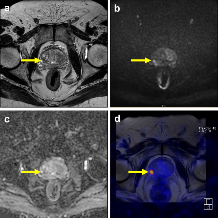
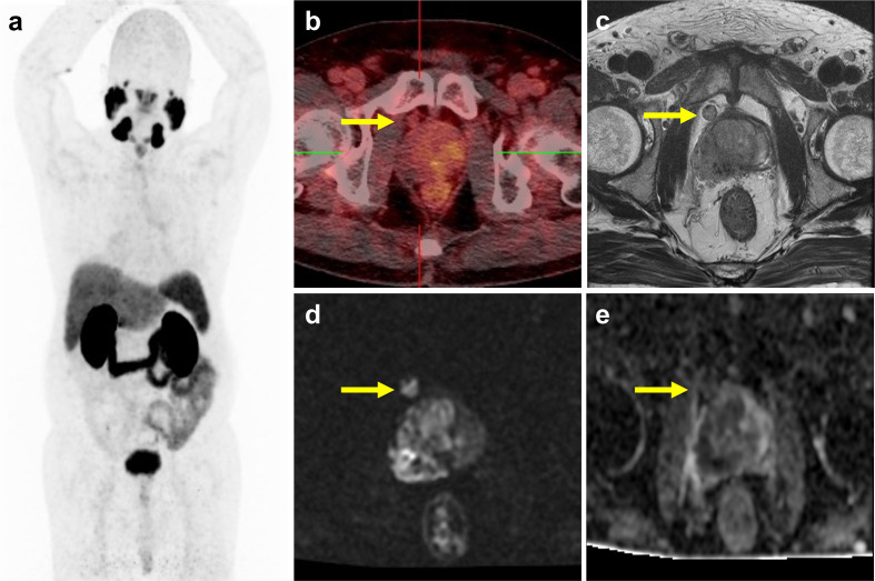
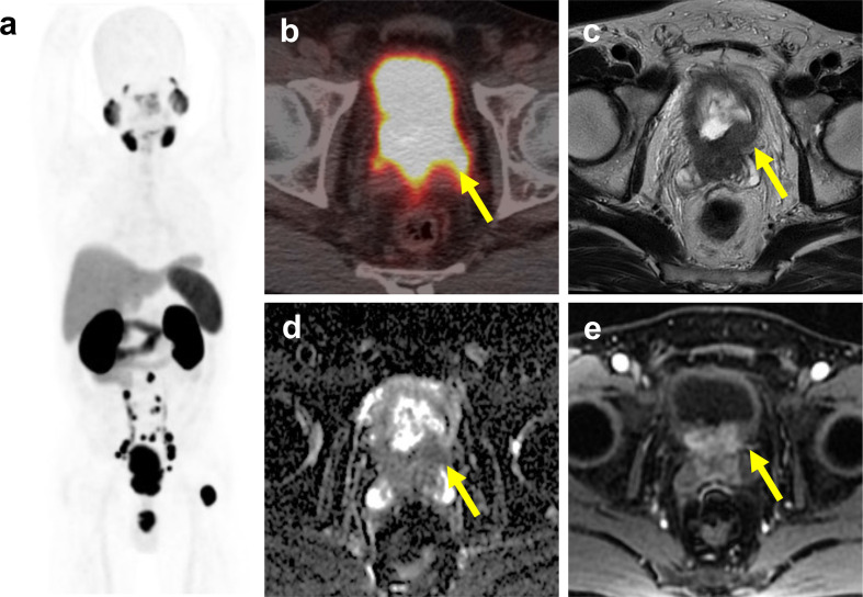
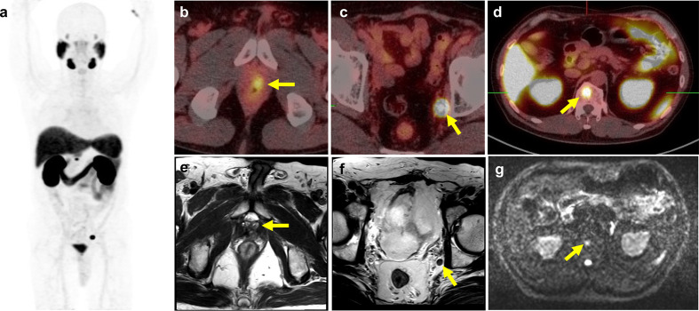
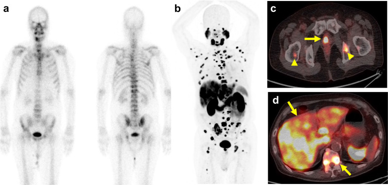
Results: We reviewed 57 original research articles during the period 2016-2021: 14 articles regarding the radiotracer PSMA; 18 articles regarding the primary tumor detection, local tumor staging, managing local recurrence; 17 articles for managing lymph node metastases; and eight articles for managing bone and other distant metastases. PSMA PET could be complementary to mpMRI for primary prostate cancer localization and is particularly valuable for PI-RADS three lesions. PET-MRI is better than PET-CT in local tumor staging due to its specific benefit in predicting extracapsular extension in MRI-occult prostate cancer patients. PET-MRI is likely superior as compared with PET-CT in detecting local recurrence, and has slightly higher detection rates than PET-CT in lymph node recurrence. PET-CT and PET-MRI seem to have equivalent performance in detecting distant bony or visceral metastases.

Conclusion: In conclusion, PET-MRI is suitable for local and regional disease, either primary staging or restaging, whereas PET-CT is valuable for managing distant bony or visceral metastasis.

Advances in knowledge: We reviewed the emerging applications of PET-MRI and PET-CT in clinical aspects. Readers will gain an objective overview on the strength and shortfalls of PET-MRI or PET-CT in the management of prostate cancer.


Figures

Figure 1.
Figure 1.
PSMA PET–MRI complementary to mpMRI for primary prostate cancer localization. A 80-year-old male with slightly increased PSA level 5.66 ng ml−1. (a) A T2 low signal intensity focus was noted at right posterolateral part of right peripheral zone, the greatest dimension was measured as 1.2 cm. (b, c) The lesion showed hyperintense on high b-value DWI and hypointense on ADC, which was considered a PIRADS four lesion. (d) The fused PET/MR image showed high 68Ga-PSMA-11 uptake at same location, and the possibility of extracapuslar extension was excluded. The final biopsy confirmed a Gleason score 4 + 3 prostate cancer lesion.
Figure 2.
Figure 2.
Low PSMA expression cancer with nodal metastasis depicted by mpMRI. A 73-year-old male with biopsy-proved prostate cancer, the Gleason score was 5 + 4, and the PSA level was 5.73 ng ml−1. (a) The PSMA maximal intensity projection did not reveal any abnormal 68Ga-PSMA-11 uptake. (b) The fused PET–CT image showed low 68Ga-PSMA-11 uptake at right portion of prostate gland and right periprostatic node, labelled by yellow arrows. (c, d, e) The MRI showed a large prostate tumor with right extracapsular extension and a periprostatic lymph node. The lesions showed restricted diffusion on DWI.
Figure 3.
Figure 3.
Prostate cancer with bladder involvement better delineated by mpMRI. A 60-year-old male with prostate cancer initial clinical stage IVB. (a) The maximal intensity projection of PSMA PET showed prostate tumor with pelvic and para-aortic lymph nodes, and left proximal femur bone metastasis. (b) The fused PET–CT image showed high 68Ga-PSMA-11 uptake at prostate gland. However, high radioactivity in the bladder limit the ability of 68Ga-PSMA PET–CT to detect bladder neck invasion by prostate tumor. Prostate cancer with bladder involvement (arrow) and seminal vesicle invasion was clearly delineated on T 2-weighted image (c), ADC map (d), and DCE MRI (e).
Figure 4.
Figure 4.
PSMA PET–MRI demonstrated local, regional and distant tumor recurrence. A 65-year-old male underwent radical prostatectomy 7 years ago, initial staging pT3bN0M0, Gleason score 4 + 3, PSA was slow raising to 4.20 ng ml−1. (a) The maximum intensity projection of PSMA PET shows abnormally-increased 68Ga-PSMA-11 uptake at (b) left prostate fossa, (c) left obturator lymph node, and (d) T12 spine. The yellow arrows on the corresponding multisequence MR imaging demonstrates (e) a focal nodule at left anastomotic site, (f) a left obturator lymph node with short axis diameter measured as 0.6 cm, and (g) a focal hyperintensity on DWI at T12 vertebral body. These lesions were considered as recurrent and metastatic prostate cancer.
Figure 5.
Figure 5.
PSMA PET–CT for managing castration-resistant prostate cancer (mCRPC). A 73-year-old male with initial staging T3N1M1b with androgen deprivation therapy since 7 years ago, PSA keep progression even after change treatment to new antiandrogen drug, castration-resistant prostate cancer (mCRPC) status was confirmed, and current PSA level was 675 ng ml−1. (a) The bone scintigraphy showed multiple hot spots involving spines, left posterior ribs, and pelvic bones. (b) The PSMA PET showed more extensively bony metastasis, (c) primary prostate cancer lesion labelled by yellow arrow, bony metastasis by arrowheads and (d) liver metastasis.

References

    1. Maurer T, Gschwend JE, Rauscher I, Souvatzoglou M, Haller B, Weirich G, et al. . Diagnostic efficacy of (68)Gallium-PSMA positron emission tomography compared to conventional imaging for lymph node staging of 130 consecutive patients with intermediate to high risk prostate cancer. J Urol 2016; 195: 1436–43. doi: 10.1016/j.juro.2015.12.025 - DOI - PubMed
    1. Afshar-Oromieh A, Babich JW, Kratochwil C, Giesel FL, Eisenhut M, Kopka K, et al. . The rise of PSMA ligands for diagnosis and therapy of prostate cancer. J Nucl Med 2016; 57(Suppl 3): 79S–89. doi: 10.2967/jnumed.115.170720 - DOI - PubMed
    1. Hoffmann MA, Miederer M, Wieler HJ, Ruf C, Jakobs FM, Schreckenberger M. Diagnostic performance of 68Gallium-PSMA-11 PET–CT to detect significant prostate cancer and comparison with 18FEC PET–CT. Oncotarget 2017; 8: 111073–83. doi: 10.18632/oncotarget.22441 - DOI - PMC - PubMed
    1. Li X, Rowe SP, Leal JP, Gorin MA, Allaf ME, Ross AE, et al. . Semiquantitative Parameters in PSMA-Targeted PET Imaging with 18F-DCFPyL: Variability in Normal-Organ Uptake. J Nucl Med 2017; 58: 942–6. doi: 10.2967/jnumed.116.179739 - DOI - PMC - PubMed
    1. Kesch C, Vinsensia M, Radtke JP, Schlemmer HP, Heller M, Ellert E, et al. . Intraindividual cComparison of 18F-PSMA-1007 PET–CT, mMultiparametric MRI, and radical prostatectomy specimens in patients with primary prostate cancer: a retrospective, proof-of-concept studyRadical Prostatectomy Specimens in Patients with Primary Prostate Cancer: A Retrospective, Proof-of-Concept Study. J Nucl Med 2017; 58: 1805–10. doi: 10.2967/jnumed.116.189233 - DOI - PubMed

MeSH terms

Substances